Fluorescence Digital Image Gallery
Normal Pig Kidney Epithelial Cells (LLC-PK1)
|
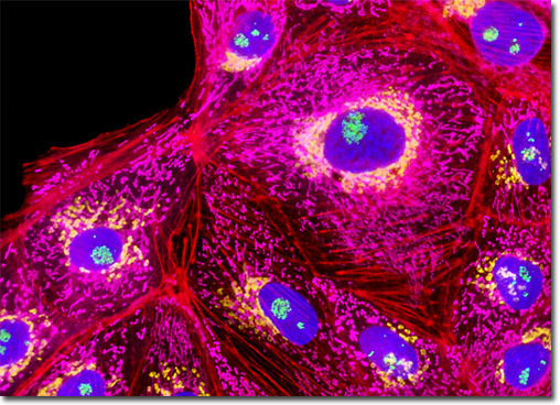
The normal pig kidney epithelial (LLC-PK1) cells presented in the digital image above were resident in an adherent culture stained for F-actin with Alexa Fluor 568 conjugated to phalloidin (orange-red fluorescence), and for DNA with the bis-benzimidazole dye Hoechst 33258 (blue fluorescence). In addition, the culture was immunofluorescently labeled with Alexa Fluor 488 (green fluorescence) and Alexa Fluor 750 (pseudocolored yellow) conjugated to goat secondary antibodies that target mouse anti-fibrillarin (nucleoli) and rabbit anti-giantin (targeting the Golgi complex) primary antibodies, respectively. The mitochondrial network was simultaneously visualized using MitoTracker Deep Red 633 (pseudocolored purple). Images were recorded in grayscale with a QImaging Retiga Fast-EXi camera system coupled to an Olympus BX-51 microscope equipped with bandpass emission fluorescence filter optical blocks provided by Omega Optical. During the processing stage, individual image channels were pseudocolored with RGB values corresponding to each of the fluorophore emission spectral profiles. |
© 1995-2025 by Michael W. Davidson and The Florida State University. All Rights Reserved. No images, graphics, software, scripts, or applets may be reproduced or used in any manner without permission from the copyright holders. Use of this website means you agree to all of the Legal Terms and Conditions set forth by the owners.
This website is maintained by our
|