Fluorescence Digital Image Gallery
Rat Jejunum Myenteric Plexus Enteroglial Cells (EGC/PK060399egfr)
|
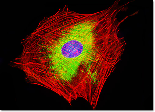
The smooth endoplasmic reticulum (ER) is the portion of the ER membrane network that does not feature ribosomes on its cytoplasmic surface. In most cells, the amount of smooth endoplasmic present is relatively small, and the membrane is primarily important as the location of exit sites where transport vesicles bud off from the ER in order to transport the proteins and lipids they contain to the Golgi complex. However, in some specialized cells, the smooth endoplasmic reticulum is significantly more extensive and carries out additional functions. For instance, cells that comprise the reproductive organs and other parts of the body that are involved in lipid metabolism are rich in smooth ER, the enzymes of which are important for the synthesis of steroids, oils, and phospholipids. Enzymes present in the smooth endoplasmic reticulum also help render drugs and poisonous substances less toxic, especially in the cells that comprise the liver. In hepatocytes, the prominent smooth ER present is particularly notable for its role in the detoxification of harmful metabolic byproducts and lipoprotein production, which is essential for the transport of lipids throughout the body via the bloodstream. The culture of EGC enteroglial cells featured in the digital image above was stained with Alexa Fluor 488 conjugated to the lectin concanavalin A, which selectively binds to alpha-mannopyranosyl and alpha-glucopyranosyl residues (primarily in the endoplasmic reticulum). Alexa Fluor 568 conjugated to phalloidin and DAPI were also used to label the culture, targeting filamentous actin and nuclear DNA, respectively. Images were recorded in grayscale with a QImaging Retiga Fast-EXi camera system coupled to an Olympus BX-51 microscope equipped with bandpass emission fluorescence filter optical blocks provided by Omega Optical. During the processing stage, individual image channels were pseudocolored with RGB values corresponding to each of the fluorophore emission spectral profiles. |
© 1995-2025 by Michael W. Davidson and The Florida State University. All Rights Reserved. No images, graphics, software, scripts, or applets may be reproduced or used in any manner without permission from the copyright holders. Use of this website means you agree to all of the Legal Terms and Conditions set forth by the owners.
This website is maintained by our
|