Fluorescence Digital Image Gallery
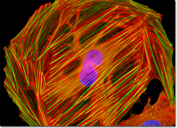
Normal African Green Monkey Kidney Fibroblast Cells (CV-1)
|
The kidney cells of African green monkeys and other primates have been heavily used to generate vaccines since the 1950s. Though CV-1 cells were not established as a research culture until the following decade, some early simian lines were found to be contaminated with certain simian viruses years after they had begun being utilized for vaccine production. Of most concern has been SV40, which was introduced to a sizeable segment of the human population through tainted polio vaccines by the time its presence in the vaccines was discovered in 1960. A Papovaviridae virus, when SV40 infects some mammals, the virus is unable to reproduce, but instead forms T-antigen and transforms host cells into cancer cells. Studies in the 1960s suggested that the virus had no effect on humans, although it was shown to cause tumor growth in laboratory animals such as hamsters. However, many investigations that have been carried out more recently indicate that SV40 may in fact be associated with certain kinds of cancers in humans, most notably mesothelioma. A consensus in the scientific community has yet to be reached, but in the meantime, cell lines used in modern vaccine manufacturing operations, including the CV-1 line, are much more stringently checked for contamination than those utilized in earlier periods. In the digital image presented above, CV-1 cells are displayed that were immunofluorescently labeled with primary anti-tubulin mouse monoclonal antibodies followed by goat anti-mouse Fab fragments conjugated to Rhodamine Red-X. In addition, the cells were stained with DAPI, which selectively binds to DNA in the cell nuclei, and Alexa Fluor 488 conjugated to phalloidin, which targets the filamentous actin network. Images were recorded in grayscale with a QImaging Retiga Fast-EXi camera system coupled to an Olympus BX-51 microscope equipped with bandpass emission fluorescence filter optical blocks provided by Omega Optical. During the processing stage, individual image channels were pseudocolored with RGB values corresponding to each of the fluorophore emission spectral profiles. |
© 1995-2025 by Michael W. Davidson and The Florida State University. All Rights Reserved. No images, graphics, software, scripts, or applets may be reproduced or used in any manner without permission from the copyright holders. Use of this website means you agree to all of the Legal Terms and Conditions set forth by the owners.
This website is maintained by our
|
