Fluorescence Digital Image Gallery
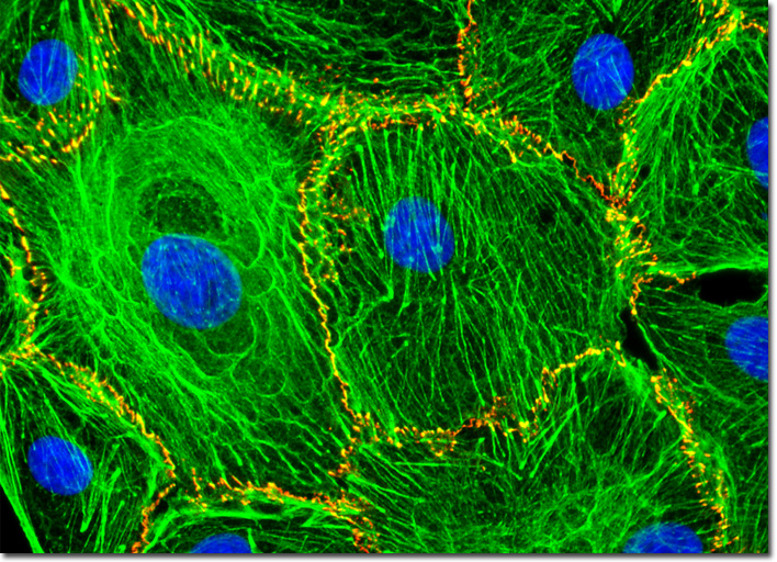
Normal African Green Monkey Kidney Fibroblast Cells (CV-1)
|
The kidney is a major excretory organ of primates and other vertebrates. The principal responsibility of the organ is to separate urea, toxins, and other types of waste from the blood, while water, salt, and electrolytes are maintained at an appropriate level. Due to this important role, the kidney is also involved in blood pressure and acid-base regulation in the body. Nephrons are the basic filtering units of the kidney, more than a million of them being present in a normal adult human kidney. Working together, the nephrons are able to filter blood at an impressive rate, processing the entire five-quart water content of the human circulatory system about every 45 minutes. Only a minute portion of the material passing through the kidneys is actually excreted, however, the vast majority being reabsorbed by the nephrons. The normal (non-transformed) African green monkey kidney fibroblast cells (CV-1 line) that are exhibited in the digital image above were immunofluorescently labeled with primary anti-cadherin monoclonal antibodies (mouse) followed by goat anti-mouse Fab fragments conjugated to Cy3. In addition, the specimen was simultaneously stained for DNA with the ultraviolet-absorbing probe DAPI, and for the cytoskeletal filamentous actin network with Alexa Fluor 488 conjugated to phalloidin. Images were recorded in grayscale with a QImaging Retiga Fast-EXi camera system coupled to an Olympus BX-51 microscope equipped with bandpass emission fluorescence filter optical blocks provided by Omega Optical. During the processing stage, individual image channels were pseudocolored with RGB values corresponding to each of the fluorophore emission spectral profiles. |
© 1995-2025 by Michael W. Davidson and The Florida State University. All Rights Reserved. No images, graphics, software, scripts, or applets may be reproduced or used in any manner without permission from the copyright holders. Use of this website means you agree to all of the Legal Terms and Conditions set forth by the owners.
This website is maintained by our
|