Fluorescence Digital Image Gallery
Mongoose Skin Fibroblast Cells (APM)
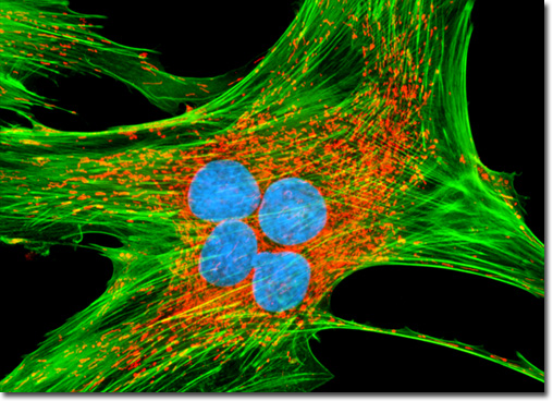
|
Mitochondria are often depicted as static, cylinder-shaped organelles, but time-lapse studies reveal that the important energy generators may change shape and move about the cytoplasm on an almost continuous basis. These activities often appear to involve the cytoskeletal microtubules, which influence the characteristic direction and dissemination of the mitochondria in various kinds of cells. The number of mitochondria present in a cell is related to the metabolic needs of that cell, and may range from a single large mitochondrion to thousands of the organelles. Their size generally ranges from 1 to 10 micrometers, making mitochondria large enough to be observed with a light microscope. The organelles were originally identified during the 1800s, and were commonly believed to transmit hereditary information until after the beginning of the twentieth century. Most of the modern understanding of the functional role of mitochondria did not develop until after a method for isolating the intact organelles was developed in 1948. The popular multiple fluorophore combination of MitoTracker Red CMXRos, Alexa Fluor 488 conjugated to phalloidin, and Hoechst 33258 was employed to perform a triple label experiment on the monolayer of African water mongoose cells presented above. The trio of fluorophores stains the cells with red (mitochondria), green (filamentous actin), and blue (DNA in the nucleus) fluorescence emission. Images were recorded in grayscale with a QImaging Retiga Fast-EXi camera system coupled to an Olympus BX-51 microscope equipped with bandpass emission fluorescence filter optical blocks provided by Omega Optical. During the processing stage, individual image channels were pseudocolored with RGB values corresponding to each of the fluorophore emission spectral profiles. |
© 1995-2025 by Michael W. Davidson and The Florida State University. All Rights Reserved. No images, graphics, software, scripts, or applets may be reproduced or used in any manner without permission from the copyright holders. Use of this website means you agree to all of the Legal Terms and Conditions set forth by the owners.
This website is maintained by our
|