Fluorescence Digital Image Gallery
Mongoose Skin Fibroblast Cells (APM)
|
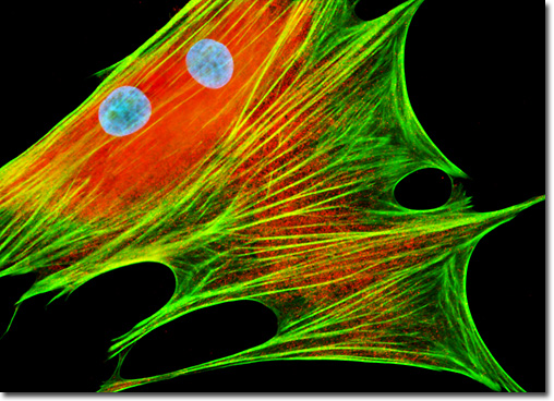
Myosin is a name utilized to describe a family of motor proteins that interact with F-actin filaments. Though a number of different myosins have been described, the most widely familiar of the proteins continues to be myosin II, the first of the family to be discovered. Each myosin II molecule present in a cell is comprised of two identical heavy chains (200 kD) and two pairs of light chains (17 to 22 kD). The enzyme trypsin can cleave the myosin heavy chains so that a rod-like tail region, called light meromyosin, and a globular head domain, termed heavy meromyosin, are formed. Light meromyosin is involved in the assembly of the bipolar thick filament, whereas heavy meromyosin is further cleaved by papain into subunits, one of which binds adenosine triphosphate (ATP) and engages in the establishment of cross-bridges between thin and thick myofilaments. Myosin II molecules are tightly packed in thick filaments (200 to 300 are present in each thick filament) and are arranged so that they are parallel, but are staggered in a manner that ensures that the middle of each filament only contains myosin tail regions and the ends of each structure are comprised of both heads and tails. A fixed and permeabilized monolayer culture of APM cells (illustrated above) was treated with mouse anti-nonmuscle myosin II (heavy chain) monoclonal primary antibodies followed by goat anti-mouse secondary antibodies (IgG) conjugated to Texas Red. Oregon Green 488 conjugated to phalloidin was included in the secondary cocktail to target the filamentous actin network. After the immunofluorescence and phallotoxin labeling, the cells were washed and counterstained with Hoechst 33258. Images were recorded in grayscale with a QImaging Retiga Fast-EXi camera system coupled to an Olympus BX-51 microscope equipped with bandpass emission fluorescence filter optical blocks provided by Omega Optical. During the processing stage, individual image channels were pseudocolored with RGB values corresponding to each of the fluorophore emission spectral profiles. |
© 1995-2025 by Michael W. Davidson and The Florida State University. All Rights Reserved. No images, graphics, software, scripts, or applets may be reproduced or used in any manner without permission from the copyright holders. Use of this website means you agree to all of the Legal Terms and Conditions set forth by the owners.
This website is maintained by our
|