Fluorescence Digital Image Gallery
Mongoose Skin Fibroblast Cells (APM)
|
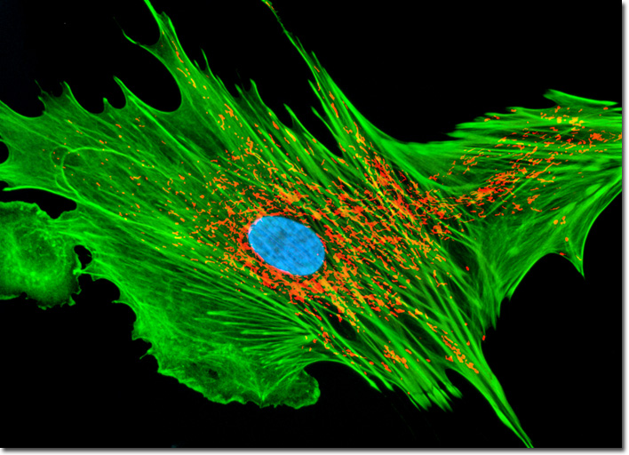
In its monomeric form, the protein actin is globular (G-actin) and generally resides throughout the cytoplasm. Cytoskeletal actin filaments, also known as microfilaments, are formed from a pair of G-actin chains that coil around one another to create an asymmetrical (with a plus and minus terminus) filamentous-form of the protein (F-actin). Some of the G-actin present in a cell does remain unpolymerized, however, due to the affinity of various small proteins for the monomeric form of actin, which inhibit polymerization by binding with specific sites on the globular protein. Actin filaments range from 5 to 9 nanometers in diameter and are designed to bear large amounts of tension. The cytoskeletal elements are generally nucleated from the minus end, which grows at a much slower pace than the positive end. When the proper length of an actin filament has been achieved, the elongation of the structure is halted through the addition of small capping proteins to the plus end of the filament. In order for a capped actin filament to resume growth, these proteins must be detached by another cellular substance, such as the phospholipid, polyphosphoinositide. Using a popular triple-fluorophore staining technique for mitochondria, filamentous actin, and the nucleus, the log phase monolayer culture of APM cells illustrated above was first treated with MitoTracker Red CMXRos for one hour, and then fixed with medium containing 3.7-percent paraformaldehyde. After permeabilization and blocking with bovine serum albumen, the cells were labeled with Alexa Fluor 488 conjugated to phalloidin and counterstained with Hoechst 33258. Images were recorded in grayscale with a QImaging Retiga Fast-EXi camera system coupled to an Olympus BX-51 microscope equipped with bandpass emission fluorescence filter optical blocks provided by Omega Optical. During the processing stage, individual image channels were pseudocolored with RGB values corresponding to each of the fluorophore emission spectral profiles. |
© 1995-2025 by Michael W. Davidson and The Florida State University. All Rights Reserved. No images, graphics, software, scripts, or applets may be reproduced or used in any manner without permission from the copyright holders. Use of this website means you agree to all of the Legal Terms and Conditions set forth by the owners.
This website is maintained by our
|