Fluorescence Digital Image Gallery
Mongoose Skin Fibroblast Cells (APM)
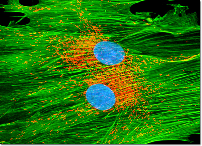
|
Two specialized membranes, both of which are phospholipid bilayers, encircle each mitochondrion present in a cell. These membranes effectively compartmentalize the organelle into a narrow intermembrane space and a much larger internal matrix, and each contains a distinctive array of proteins that are associated with their functions. The outer membrane of a mitochondrion, which acts similar to a sieve, includes numerous copies of the channel-forming protein porin and is impenetrable to molecules greater than 5000 Daltons in size. Most of the molecules that are small enough to pass through the outer membrane are impeded by the more-selective inner membrane that is typically highly convoluted so that a large number of infoldings called cristae are formed. The inner membrane contains an array of transport proteins that render it selectively permeable to molecules that need to move into and out of the matrix, and also possesses a large amount of the phospholipid cardiolipin, which makes the lipid bilayer particularly difficult for ions to penetrate. In addition, proteins embedded in the inner mitochondrial membrane are essential for oxidation reactions and the generation of adenosine triphosphate (ATP) in the internal matrix. Applying a collection of popular mitochondrial, actin, and DNA probes, the culture of APM cells presented above was grown to log phase, treated with MitoTracker Red CMXRos before fixing, and then labeled with phalloidin and Hoechst 33258 after permeabilization. The red fluorescence arises from the mitochondrial dye (MitoTracker), while the phalloidin was conjugated to Alexa Fluor 488 to generate green fluorescence. Nuclei are rendered in light blue. Images were recorded in grayscale with a QImaging Retiga Fast-EXi camera system coupled to an Olympus BX-51 microscope equipped with bandpass emission fluorescence filter optical blocks provided by Omega Optical. During the processing stage, individual image channels were pseudocolored with RGB values corresponding to each of the fluorophore emission spectral profiles. |
© 1995-2025 by Michael W. Davidson and The Florida State University. All Rights Reserved. No images, graphics, software, scripts, or applets may be reproduced or used in any manner without permission from the copyright holders. Use of this website means you agree to all of the Legal Terms and Conditions set forth by the owners.
This website is maintained by our
|