Fluorescence Digital Image Gallery
Mongoose Skin Fibroblast Cells (APM)
|
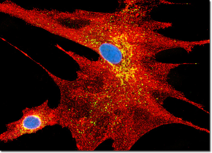
The protein clathrin is composed of three heavy chains and three light chains that self-assemble into a triskelion, a structure with three bent legs radiating from a central point. These clathrin triskelions form polyhedral lattices, termed clathrin baskets or coats, on the cytosolic surface of the plasma membrane and the trans face of the Golgi complex. The precise mechanisms involved in the assembly of clathrin baskets remain to be discovered, as are the exact mechanisms associated with the removal of the lattice from budded vesicles. Research strongly suggests, however, that clathrin coats are involved in the process of protein sorting and that a chaperone protein from the hsp70 family acts as an uncoating ATPase, utilizing energy generated by ATP hydrolysis to remove the clathrin basket. The protein auxillin that can be found attached to clathrin-coated vesicles is thought to trigger this action. The culture of APM cells featured above was immunofluorescently labeled with primary anti-clathrin (heavy chain) mouse monoclonal antibodies followed by goat anti-mouse Fab fragments conjugated to Texas Red in order to target the cytoskeletal network. In addition, peroxisomes present in the culture were simultaneously labeled with Oregon Green 488 conjugated to goat secondary antibodies directed against rabbit anti-PMP 70 (peroxisomal membrane protein 70) primary antibodies. Nuclei were counterstained with Hoechst 33258. Images were recorded in grayscale with a QImaging Retiga Fast-EXi camera system coupled to an Olympus BX-51 microscope equipped with bandpass emission fluorescence filter optical blocks provided by Omega Optical. During the processing stage, individual image channels were pseudocolored with RGB values corresponding to each of the fluorophore emission spectral profiles. |
© 1995-2025 by Michael W. Davidson and The Florida State University. All Rights Reserved. No images, graphics, software, scripts, or applets may be reproduced or used in any manner without permission from the copyright holders. Use of this website means you agree to all of the Legal Terms and Conditions set forth by the owners.
This website is maintained by our
|