Fluorescence Digital Image Gallery
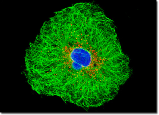
Human Lung Carcinoma Cells (A-549)
|
A carcinoma, such as the one that served as the original source of the A-549 cell line, is a solid, cancerous growth that begins its development in the epithelial tissue that forms human skin and comprises the linings of most organs and glands. The cells of a carcinoma may spread, however, to nearby tissues that are otherwise healthy and can also generate secondary tumors known as metastases in parts of the body that are distant from the initial growth. Some of the most common areas of carcinoma development include the skin, lungs, uterus, stomach, ovaries, and prostate, though the incidence of growths in these and other areas vary significantly by country. Lung carcinoma, which was deemed relatively rare in the early twentieth century, is currently diagnosed more than any other form of major cancer worldwide. It is also the most common cause of cancer fatalities in both men and women. In countries where cigarette smoking has been prevalent for many years, as many as 90 percent of patients diagnosed with lung cancer are, or have been, smokers. The human lung carcinoma cell presented in the digital image above was resident in a culture immunofluorescently labeled with primary anti-cytokeratin (pan) mouse monoclonal antibodies followed by goat anti-mouse Fab fragments conjugated to Cy2. In addition, the specimen was stained with MitoTracker Red CMXRos and Hoechst 33258 to label the mitochondrial network and nuclei, respectively. Images were recorded in grayscale with a QImaging Retiga Fast-EXi camera system coupled to an Olympus BX-51 microscope equipped with bandpass emission fluorescence filter optical blocks provided by Omega Optical. During the processing stage, individual image channels were pseudocolored with RGB values corresponding to each of the fluorophore emission spectral profiles. |
© 1995-2025 by Michael W. Davidson and The Florida State University. All Rights Reserved. No images, graphics, software, scripts, or applets may be reproduced or used in any manner without permission from the copyright holders. Use of this website means you agree to all of the Legal Terms and Conditions set forth by the owners.
This website is maintained by our
|
