Fluorescence Digital Image Gallery
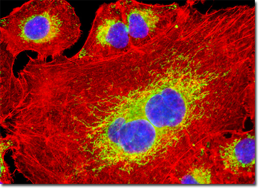
Human Lung Carcinoma Cells (A-549)
|
Lung carcinomas are not usually detected at an early stage of their development because symptoms of their presence, which may include chest pain, difficulty breathing, persistent coughing, bloody phlegm, and increased susceptibility to respiratory infections, do not usually appear until they are in an advanced state. Oftentimes, the cancerous growths are not noticed until they have spread to other locations within the body. Thus, the prognosis for patients with lung cancer is somewhat bleak, the five-year survival rate for them being estimated at less than 15 percent overall. Treatments for the disease are similar to those for other types of cancers, consisting primarily of chemotherapy, surgery, and radiation exposure. The exact treatment chosen depends upon a number of factors, such as the patient’s health and the level of advancement of the disease. In some instances, for example, it may be judged necessary to remove part or all of a lung, while in other cases, radiation therapy to destroy small numbers of cancer cells may be judged sufficient. Frequently, the various treatments will be used in conjunction with one another. The adherent culture of A-549 cells displayed in the digital image above was stained for F-actin with Alexa Fluor 568 conjugated to phalloidin, and for DNA with 4',6-diamidino-2-phenylindole (DAPI). The cells were additionally labeled for lipid vesicles with Alexa Fluor 488 conjugated to lectin PNA, a peanut-derived protein that binds to specific carbohydrate groups on proteins or cell membranes. Images were recorded in grayscale with a QImaging Retiga Fast-EXi camera system coupled to an Olympus BX-51 microscope equipped with bandpass emission fluorescence filter optical blocks provided by Omega Optical. During the processing stage, individual image channels were pseudocolored with RGB values corresponding to each of the fluorophore emission spectral profiles. |
© 1995-2025 by Michael W. Davidson and The Florida State University. All Rights Reserved. No images, graphics, software, scripts, or applets may be reproduced or used in any manner without permission from the copyright holders. Use of this website means you agree to all of the Legal Terms and Conditions set forth by the owners.
This website is maintained by our
|