Fluorescence Digital Image Gallery
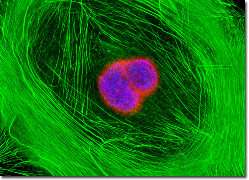
Embryonic Swiss Mouse Fibroblast Cells (3T3)
|
In eukaryotes, the nuclear envelope encloses the chromosomes (DNA) and other organelles, such as the nucleoli, to clearly define the nucleus of the cell. The envelope is comprised of two distinct concentric membranes that contain perforations in regions surrounded by a family of proteins known as the nuclear pore complexes. Each pore complex contains more than 50 different integral proteins (termed nucleoporins) that have a combined molecular weight of approximately 125 megaDaltons. The nucleoporins are geometrically arranged in a very elaborate octagonal symmetry, which has been extensively studied using optical and electron microscopy. In fluorescence microscopy, the nuclear pore complexes can be visualized using antibodies targeting any of the nucleoporin subfamilies to investigate the morphology and composition of the nucleus and nuclear envelope. Using a monoclonal antibody directed against a related family of nuclear pore complex proteins, the adherent culture of Swiss mouse embryo cells presented above was imaged after being fixed, permeabilized, and treated with primary mouse antibodies followed by goat anti-mouse secondary antibodies conjugated to Alexa Fluor 568 (red fluorescence). The cells were subsequently counterstained with Alexa Fluor 488 conjugated to phalloidin (filamentous actin; green fluorescence) and Hoechst 33342 (DNA in the nucleus; blue fluorescence). Images were recorded in grayscale with a QImaging Retiga Fast-EXi camera system coupled to an Olympus BX-51 microscope equipped with bandpass emission fluorescence filter optical blocks provided by Omega Optical. During the processing stage, individual image channels were pseudocolored with RGB values corresponding to each of the fluorophore emission spectral profiles. |
© 1995-2025 by Michael W. Davidson and The Florida State University. All Rights Reserved. No images, graphics, software, scripts, or applets may be reproduced or used in any manner without permission from the copyright holders. Use of this website means you agree to all of the Legal Terms and Conditions set forth by the owners.
This website is maintained by our
|