Fluorescence Digital Image Gallery
Embryonic Swiss Mouse Fibroblast Cells (3T3)
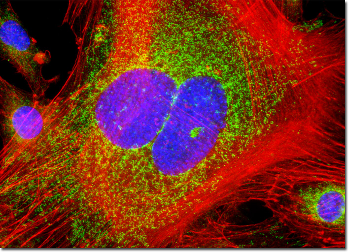
|
Soon after the 3T3 line was introduced, the scientists that developed the line noticed that cytoplasmic lipids sometimes accumulated on cultures of the fibroblasts. However, a decade passed by before Howard Green began to investigate the phenomenon in earnest. His studies on the topic seemed to suggest that although 3T3 cells are fibroblasts, they also enjoy a latent course of differentiation that converts the cells to adipocytes under certain conditions. Despite strong evidence of this adipose differentiation capability of 3T3 cells, the idea of such activity in a fibroblast line was viewed with significant skepticism. Only when Green and associates demonstrated in the late 1970s that pre-adipose 3T3 cells injected into athymic mice developed into mature pads of fat did the concept begin to gain widespread acceptance. The 3T3 cells that are featured in the digital image above were residents of a log phase culture that was immunofluorescently labeled with primary anti-human golgin-97 mouse monoclonal antibodies followed by goat anti-mouse Fab fragments conjugated to Alexa Fluor 488, in order to target the Golgi apparatus. In addition, the cells were labeled with Alexa Fluor 568 conjugated to phalloidin for filamentous actin and Hoechst 33258 for DNA in cell nuclei. Images were recorded in grayscale with a QImaging Retiga Fast-EXi camera system coupled to an Olympus BX-51 microscope equipped with bandpass emission fluorescence filter optical blocks provided by Omega Optical. During the processing stage, individual image channels were pseudocolored with RGB values corresponding to each of the fluorophore emission spectral profiles. |
© 1995-2025 by Michael W. Davidson and The Florida State University. All Rights Reserved. No images, graphics, software, scripts, or applets may be reproduced or used in any manner without permission from the copyright holders. Use of this website means you agree to all of the Legal Terms and Conditions set forth by the owners.
This website is maintained by our
|