Featured Microscopist: Spike (M.I.) Walker

Ciliate Protozoan

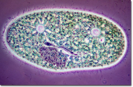
Paramecium bursaria

Phase contrast photomicrograph of Paramecium bursaria. A living specimen of this ciliate protozoan was trapped under a cover glass and slightly compressed by carefully removing water from the edge of the cover with a strip of filter paper. Note the hair-like cilia with which it swims, and also the green symbiotic cells of Zoöchlorella. Careful observation of the photomicrograph reveals a macronucleus with micronucleus in a notch, the protozoan's gullet (grey line in front of the micronucleus), and two contractile vacuoles with radiating feeder canals. The objective was a 40x/0.65 NA achromat with a Reichert Zetopan microscope. The camera system was a Reichert Zetopan with an Exacta camera body. A Zeiss microflash illumination was utilized to make the 0.5 micro second exposure. The film was Kodachrome 25. (125x)


Text and graphics for this article are
© 2000-2022 by Spike (M. I.) Walker.
All Rights Reserved under copyright law.
© 1995-2022 by Michael W. Davidson and The Florida State University. All Rights Reserved. No images, graphics, software, scripts, or applets may be reproduced or used in any manner without permission from the copyright holders. Use of this website means you agree to all of the Legal Terms and Conditions set forth by the owners.
This website is maintained by our
Graphics & Web Programming Team
in collaboration with Optical Microscopy at the
National High Magnetic Field Laboratory.
Last modification: Monday, Dec 01, 2003 at 12:54 PM
Access Count Since November 18, 2000: 11045