
Polarized Light Microscopy Digital Image Gallery
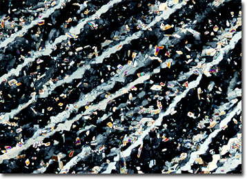
Urea
Urea is an organic compound that is the primary nitrogen-containing end product of protein metabolism in mammals, as well as some varieties of fish. The colorless substance was first isolated by French scientist Hilaire-Marin Rouelle in 1773, making it the first animal metabolite to be isolated in the form of crystals.
In 1828, German chemist Friedrich Wohler prepared urea from ammonium cyanate, a feat generally considered the earliest occurrence of an organic compound synthesis achieved through the use of inorganic materials. Since that time, the synthesis of urea has developed into a significant commercial endeavor, tremendous amounts of the substance being produced each year for use in a variety of products. The greatest use of urea is as a fertilizer and feed supplement, but the diamide of carbonic acid, sometimes referred to as carbamide, is also often utilized as a base material for the industrial production of pharmaceuticals and plastics and as a stabilizer in certain types of explosives.
Although the greatest concentration of urea within the body is found in the urine, the nitrogenous substance also occurs in the blood, bile, and perspiration, as well as the milk of lactating females. The substance is periodically excreted in the urine, but reaccumulates during protein metabolism, when amino groups (NH2) are removed from the amino acids of proteins and are converted to ammonia (NH3). Since ammonia is toxic, the body quickly converts the compound to urea by filtering it through the liver. This urea is then transferred to the kidneys via the blood, where it is prepared for excretion.