
Polarized Light Microscopy Digital Image Gallery
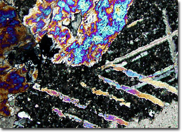
Tactite Skarn
Tactite is a type of rock that exhibits a complex mineralogical composition. Generally considered a type of skarn, tactite is formed by contact metamorphism and metasomatism of carbonate rocks.
View a second image of Tactite Skarn
Coined in Sweden as a term for deposits of iron that were found in rocks near a pluton, today skarn is more broadly understood to refer to any metamorphic zone that develops around contact areas nearby igneous rock intrusions. These zones are characterized by the introduction of large amounts of iron, silicon, aluminum, and magnesium into limestones and other carbonate sedimentary rocks. In addition to tactite, skarns are known by a number of other names, such as igneous-metamorphic and pyrometasomatic deposits.
Skarns can be found around the world and most developed during the Mesozoic period some 63 million to 230 million years ago. Zones of skarn are usually relatively irregular in shape due to the erratic characteristics of the host rocks, which vary in porosity, permeability, and lithology. The zones often contain deposits of iron, copper, gold, magnetite, tungsten, and other ores, though in relatively small amounts. Nevertheless, the small deposits are often mined for commercial profit. Some of the better-known ore-containing skarns can be found in Cornwall, Pennsylvania, Pine Creek, California, and the Copper Canyon of Nevada.