Microscopy Primer
Light and Color
Microscope Basics
Special Techniques
Digital Imaging
Confocal Microscopy
Live-Cell Imaging
Photomicrography
Microscopy Museum
Virtual Microscopy
Fluorescence
Web Resources
License Info
Image Use
Custom Photos
Partners
Site Info
Contact Us
Publications
Home

The Galleries:

Photo Gallery
Silicon Zoo
Pharmaceuticals
Chip Shots
Phytochemicals
DNA Gallery
Microscapes
Vitamins
Amino Acids
Birthstones
Religion Collection
Pesticides
BeerShots
Cocktail Collection
Screen Savers
Win Wallpaper
Mac Wallpaper
Movie Gallery

Polarized Light Microscopy Digital Image Gallery
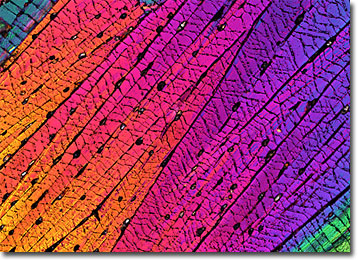
Rhodamine

A rhodamine is any of a group of orange, red, and pink dyes that are utilized for a variety of purposes. Synthesized in the laboratory, rhodamines are produced from phthalic anhydride and aminophenols.

View a second image of Rhodamine

Many rhodamines are fluorescent and are heavily utilized as fluorochromes in the field of microscopy. The dyes are especially well suited for labeling proteins and membranes, but can be used for other applications as well. Some rhodamines, for instance, such as lissamine rhodamine, gain wide use in flow tracing studies, while others, like tetramethyl rhodamine, are more popular in energy transfer research. Also, rhodamine 6G, which produces an orange-yellow light, can be utilized as a dye laser.

In addition to scientific applications, rhodamines are often useful in ways that make them more familiar to a broader public. For example, some varieties of rhodamine are utilized in the textile industry and may be responsible for the pinkish hue of a silk shirt or the red coloring of a wool coat. Rhodamines are also often added to outdoor paints used for billboards and other forms of advertising. The strong fluorescing of the molecules in the dyes makes the signs easier to see, especially at night. Rhodamines do, however, exhibit a relatively poor stability in light, although the dyes are significantly more photostable than fluorescein dyes.


BACK TO THE CHEMICAL CRYSTALS GALLERY

BACK TO THE POLARIZED LIGHT GALLERY

Questions or comments? Send us an email.
© 1998-2022 by Michael W. Davidson and The Florida State University. All Rights Reserved. No images, graphics, scripts, or applets may be reproduced or used in any manner without permission from the copyright holders. Use of this website means you agree to all of the Legal Terms and Conditions set forth by the owners.
This website is maintained by our
Graphics & Web Programming Team
in collaboration with Optical Microscopy at the
National High Magnetic Field Laboratory.
Last modification: Friday, Nov 13, 2015 at 02:19 PM
Access Count Since January 23, 2004: 13064
All of the images in this gallery were captured with a QImaging Retiga camera system.
For more information on these cameras, use the button below to access
the QImaging website:
Visit the QImaging website.
For more information on microscope manufacturers,
use the buttons below to navigate to their websites: