
Polarized Light Microscopy Digital Image Gallery
Ramie Fiber
Ramie is a vegetable fiber that has been exploited for human use for thousands of years. Naturally white and glossy, fabrics made from ramie fibers have a beautiful, lustrous appearance.
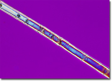
Native to Southeast Asia, the plant from which ramie fiber, also known as China grass, is derived is a member of the nettle family scientifically described as Boehmeria nivea. The extraction of fibers from the plant can be a somewhat complex and expensive process because several steps are required in order to successfully separate the filaments from surrounding gums and resins. The spinning and weaving of ramie can also be an intricate and difficult endeavor due to the characteristic brittleness and relative inflexibility of the fibers. Such attributes have significantly limited the use of ramie in commercial products.
The greatest amount of ramie production takes place in China, but Brazil, Korea, and the Philippines are also major producers. Extremely absorbent and strong, when utilized alone, ramie material dries quickly, resists shrinkage, and deters mildew, mold, and insect attacks. Its negative characteristics, however, make the fibers much better suited for fabric blends. For instance, when ramie is combined with cotton, it increases the strength, color, and luster of the fabric without excessively decreasing its elasticity. Ramie blends are therefore utilized in a variety of products, including apparel, upholstery, and netting.