
Polarized Light Microscopy Digital Image Gallery
Mylonite
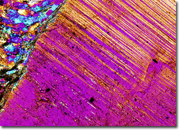
The term mylonite was first utilized in 1885 by Charles Lapworth to describe a fine-grained, laminated rock that he discovered in the Moine Thrust Zone of Scotland. Since that time, the word has become a general structural indicator of any distinctly foliated, fine-grained rock associated with a high level of ductile deformation.
View a second image of Mylonite
Mylonites, which can be found among rocks of all ages, are typically believed to be only formed in shear zones. Pressure, temperature, and strain are the primary factors that determine whether or not a mylonite or some other type of metamorphic rock will form when intense folding or faulting occurs. If these dynamics fall outside of a certain range, the process of metamorphism may result, for instance, in a cataclastite, gneiss, or granulite, rather than a fine-grained, highly laminated mylonite. In addition, mylonites require two discrete kinds of constituents in order to form: a hard element that generates larger-grained porphyroclasts and a softer element, which develops into the rock’s fine-grained matrix.
Zones of mylonite occur around the world and vary greatly in size. In Nevada, deposits of mylonite have been found that are not more than a centimeter thick, while in Canada, a mylonite zone exists that is several kilometers thick and several hundred kilometers long. No matter their size, deposits of mylonite can reveal important clues regarding the geological history of the region in which they are found. The rocks are, for example, clear indicators of the presence of a shear zone that was previously active, and close examination of samples may bring to light details concerning the type and extent of shearing that transpired.