
Polarized Light Microscopy Digital Image Gallery
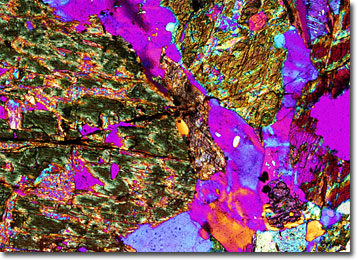
Augen Gneiss
A medium- to coarse-grained rock, gneiss is primarily distinguished by its characteristic banding caused by the segregation of its mineral constituents. Unlike schist, however, gneiss does not readily cleave along these parallel layers.
View a second image of Augen Gneiss
Gneiss is difficult to describe because it varies to such a wide extent. Nevertheless, generally the rock contains relatively large amounts of feldspar and quartz, as well as mica or some other dark, rock-forming mineral. The bands of the rock, which is formed via high-grade regional metamorphism, may be nearly parallel to the ground or steeply angled. The precise orientation of the bands may be in some way determined by characteristics of the parent rock or the type of stresses undergone during metamorphism.
Gneisses are classified based upon a variety of characteristics, such as constituent minerals, parent materials, and chemical composition. Some, such as augen gneiss, are also described based upon their structure. This type of gneiss gains its name from the elliptic or lens-shaped form of many of its mineral grains. Indeed, the word augen is German for “eye,” a reference to the appearance of these readily visible components of metamorphic rocks. To some people, an augen structure appears more akin to nuts than eyes. In fact, the scenic Hickory Nut Gorge in North Carolina is believed to have been named for the nut-shaped minerals found in much of the exposed rock present in that location.