
Phase Contrast Image Gallery
Pulmonary Emphysema
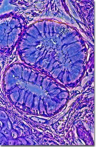
A stained thin section of human lung tissue exhibiting damage from pulmonary emphysema is illustrated below. As evidenced by the micrograph, combining phase contrast microscopy with classical histological staining techniques in pathological research often yields enhancement of cellular features.
Emphysema is one of the most common and crippling of respiratory diseases, and it is incurable. Along with chronic bronchitis and asthma, it is the 4th leading cause of death in the United States, killing over 25,000 people a year. Currently, about 2 million in the U.S. are living with emphysema.
The condition occurs when elastic fibers in the air sacs (alveoli) of the lungs are destroyed, causing the alveoli to become over-stretched. Once these sacs lose their elasticity, they are unable to push air out, or exhale. Pockets of "dead air," or carbon dioxide, become trapped in the lungs, decreasing the amount of oxygen that is being transferred to the bloodstream.
Emphysema doesn't develop suddenly, most often taking years to develop. The causes are not well understood yet, but over 80% of cases are associated with cigarette smoking. Other factors causing lung damage can lead to emphysema as well, such as asthma, tuberculosis, and exposure to heavily polluted air and dusts (e.g. coal dust).
One form of emphysema is caused by a genetic disorder and begins to affect people when they are in their 30s and 40s. It is estimated that 50,000 to 100,000 people in the U.S. were born with a deficiency of a protein known as alpha 1-antitrypsin (AAT) which can lead to an inherited form of emphysema called alpha 1-antitrypsin (AAT) deficiency-related emphysema (A1AD). AAT protects the lungs from a natural enzyme, neutrophil elastase, which helps fight bacteria and clean up dead lung tissue. Without the genetic information that tells the body how to produce AAT, the lung tissues are increasingly damaged by the activity of the enzyme.
BACK TO THE PHASE CONTRAST GALLERY