
Phase Contrast Image Gallery
Hydatid Disease
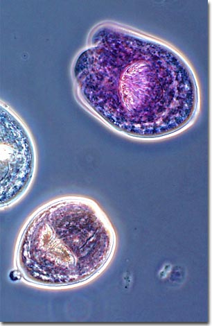
Also called echinococcal disease or hydatidosis, hydatid disease is caused by the larval form of the tapeworm parasite, Echinococcus granulosus. The photomicrograph presented below is a phase contrast image of these larvae.
The adult Echinococcus tapeworm parasitizes dogs and other canines, but requires an intermediate host (sheep, cattle, goats, wild ruminants, pigs, or humans) to complete its life cycle. The genus has a worldwide distribution, but there is considerable variation amongst the species, with differing preferences for intermediate hosts.
Dogs become infected when they eat raw or undercooked offal (especially liver, lungs, or brain) that contains hydatid cysts. The cysts are filled with protoscolices (singular: protoscolex), the larval form of the tapeworm. After being eaten, the cyst wall is digested, freeing the larvae into the intestines where they attach and grow into adult tapeworms. The adults produce eggs, which are passed to the outside with fecal material.
The eggs are ingested by the intermediate host (generally sheep or cattle) when it grazes on grass contaminated with feces from the canine host. The eggs hatch in the intestine, penetrate the gut wall, and travel via the lymphatic or blood system throughout the body where they will lodge in tissues and develop into cysts. The cysts can develop anywhere, but the majority of the cysts are found in the liver, lungs, brain, and kidneys. It takes one to two years for the cysts to produce infective protoscolices.
Humans become infected after inadvertently ingesting eggs, usually after being licked on the mouth by an tapeworm infested dog after it's been cleaning itself, or through poor hygiene practices after handling dog feces. While some people may have cysts in their bodies 5-20 years without experiencing symptoms, or may never experience symptoms, some cysts can grow large enough to cause serious health problems, especially if one or more cysts develop in the brain.
BACK TO THE PHASE CONTRAST GALLERY