Microscopy Primer
Light and Color
Microscope Basics
Special Techniques
Digital Imaging
Confocal Microscopy
Live-Cell Imaging
Photomicrography
Microscopy Museum
Virtual Microscopy
Fluorescence
Web Resources
License Info
Image Use
Custom Photos
Partners
Site Info
Contact Us
Publications
Home

The Galleries:

Photo Gallery
Silicon Zoo
Pharmaceuticals
Chip Shots
Phytochemicals
DNA Gallery
Microscapes
Vitamins
Amino Acids
Birthstones
Religion Collection
Pesticides
BeerShots
Cocktail Collection
Screen Savers
Win Wallpaper
Mac Wallpaper
Movie Gallery

Phase Contrast Image Gallery

Fish Cycloid Scale

A stained thin section of fish cycloid scale, illustrated below, was photographed using phase contrast optics. The image was captured using an Olympus IX-70 inverted microscope equipped with a DP-10 digital camera.

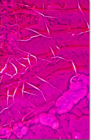
Cycloid scales are smooth, flat, and round with concentric lines called circuli. This type of overlapping scale demonstrates added flexibility and grows larger as the fish grows. In some species, cycloid scales possess annual growth rings. Scales, divided into several categories on the basis of composition and structure, provide protection from the environment and predators and are found on almost all fish. Fish scales are formed from skeletal elements (bone) that emerge from the dermal layer of skin. Other types of scales include bony, spiny projections with an enamel-like covering termed placoid scales. Another group of scales is similar to placoid scales, but are covered with an enamel-like substance called ganolin. Many scientists believe that teeth evolved from placoid scales. Cycloid scales are found on advanced fish species such as trout, herring, and carp.

BACK TO THE PHASE CONTRAST GALLERY

Questions or comments? Send us an email.
© 1998-2022 by Michael W. Davidson and The Florida State University. All Rights Reserved. No images, graphics, scripts, or applets may be reproduced or used in any manner without permission from the copyright holders. Use of this website means you agree to all of the Legal Terms and Conditions set forth by the owners.
This website is maintained by our
Graphics & Web Programming Team
in collaboration with Optical Microscopy at the
National High Magnetic Field Laboratory.
Last modification: Friday, Nov 13, 2015 at 02:19 PM
Access Count Since April 23, 2000: 28519
For more information on microscope manufacturers,
use the buttons below to navigate to their websites: