
Phase Contrast Image Gallery
Chronic Pneumonia
A stained thin section of human lung tissue exhibiting damage from chronic pneumonia is illustrated below. As evidenced by the micrograph, combining phase contrast microscopy with classical histological staining techniques in pathological research often yields enhancement of cellular features.
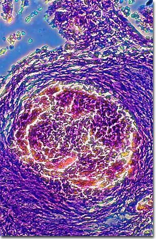
Pneumonia is an inflammation of the lungs that can be caused by one of any number of agents or microorganisms. Chronic pneumonia, also known as Loeffler's syndrome or eosinophilic pneumonia, is a rare disorder that is considered to be the result of an allergic reaction or reaction to parasites.
Eosinophils are one of five white blood cell types and specifically act to protect the body during allergic reactions, moderating its own defenses. During the body's reaction to a foreign body, eosinophils are produced in greater numbers, engulfing antigen-antibody complexes and limiting the effects of certain chemicals the body produces in its defense, such as histamine. Like other white blood cells, eosinophils are nucleated and capable of amoeba-like movement.
In Loeffler's syndrome, lung alveoli (the lung's tiny air sacs) fill with fluid and eosinophils, causing the symptoms of pneumonia. The condition usually resolves itself without treatment, but sometimes the symptoms are severe enough to warrant treatment.
BACK TO THE PHASE CONTRAST GALLERY