
Phase Contrast Image Gallery
Camarasaurus: Chambered Lizard
Thin sections of fossilized dinosaur bones often reveal clues about the anatomy and physiology of these long-extinct, but fascinating creatures. The photomicrograph below illustrates a phase contrast image of a 30-micron thick thin section of a camarasaurus bone.
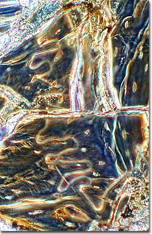
Analysis of this photomicrograph reveals an agate-like deposition of minerals within the canals of the bone structure. These deposits are often birefringent and can display a kaleidoscopic array of color when viewed under polarized illumination with a microscope.
Camarasaurs were dinosaurs commonly found in North America during the late Jurassic period, 163 to 144 million years ago. They belonged to the Sauropod family of dinosaurs, the largest animals that ever lived on land, which included the well-known Apatosaurus (formerly known as Brontosaurus). Sauropods were quadrupedal herbivores and ranged in size from 21 to 120 feet in length (7-40 meters).
Camarasaurus is the only sauropod for which a complete skeleton has been found. These long-necked dinosaurs were somewhat smaller than other sauropods of the time and grew to a length of about 59 feet (18 meters). Camarasaurs were further distinguished by their shorter necks and tails; higher, box-like skulls; and large, spoon-shaped teeth. Huge weight-saving hollows in its vertebrae gave this sauropod its name, "chambered lizard".
BACK TO THE PHASE CONTRAST GALLERY