
Fluorescence Digital Image Gallery
Wheat Rust Pustule
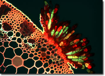
The photomicrograph below is a fluorescence image of a stained thin section of wheat showing a brick-red pustule captured erupting from the leaf surface. Wheat rust is a common and serious disease, reducing crop yields both in the United States and in other wheat-growing areas of the world.
Wheat rust is caused by a parasitic fungus and can affect both the leaves and stems of wheat plants. In individual fields, the disease accounts for crop losses ranging from trace amounts to as much as 40 percent when weather conditions are favorable for fungus growth. Rust infections usually appear as yellow, orange, red, rust, brown, or black powdery pustules on leaves, young shoots, and fruits.
The fungus affecting the plants produces two types of fruiting bodies during its life cycle, aecia and pycnia. These structures produce different types of spores, aeciospores and pycniospores, which are transmitted from one plant to another by the wind. The pycniospores parasitize wheat while the aeciospores can infect other plants.
The specimen presented here was imaged with a Nikon E600 microscope operating with fluorite and/or apochromatic objectives and vertical illuminator equipped with a mercury arc lamp. Specimens were illuminated through Nikon dichromatic filter blocks containing interference filters and a dichroic mirror and imaged with standard epi-fluorescence techniques. Specific filters for the wheat rust thin section were a B-2E/C and a Y-2E/C. Photomicrographs were captured with a Optronics MagnaFire digital camera system coupled to the microscope with a lens-free C-mount adapter.
VIEW THIS SPECIMEN UNDER PHASE CONTRAST ILLUMINATION
BACK TO THE FLUORESCENCE DIGITAL IMAGE GALLERY