
Fluorescence Digital Image Gallery
Pine Needle Cross Section
The slender, green needles characteristic of pine trees may not look like leaves, but they are. In fact, the streamlined projections are brilliantly designed so that they can convert sunlight into food even on the coldest days of winter, while minimizing the amount of evapotranspiration that takes place during the heat of the summer.
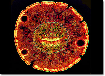
Due to the efficiency of their needles, pine trees remain covered in leaves year-round and are commonly known as evergreens. Older needles drop off periodically (usually when they are two to four years old), but they are continuously replaced with new growth. The needles grow in sheathed bundles, arranged spirally along supporting shoots. The number of needles in each bundle may vary, but they generally contain two to three needles on hard pines and five on soft pines.
While pine trees are most often commercially exploited for their wood, human use of their needles has been ongoing for thousands of years. Native Americans frequently utilized the youngest leaves of pines to brew a mild red tea used for medicinal purposes. They also gathered fallen needles from the ground in order to weave baskets, a practice that continues in modern times. Another contemporary method of exploiting pine needles involves extracting their essential oil through distillation. The fragrant substance, which some argue has antimicrobial, antiviral, and antiseptic properties, is often used in massage therapy.
BACK TO THE FLUORESCENCE DIGITAL IMAGE GALLERY