
Fluorescence Digital Image Gallery
Moss Reproductive Tissue
Mosses are the most common, diverse, and advanced group of bryophytes, a division of green, seedless plants that dates back to the Permian period (286 to 245 million years ago). In bryophytes, the antheridium is the male sex organ, which produces sperm. The archegonium, illustrated along with the antheridium in the fluorescence photomicrograph presented below, is the female reproductive organ, which produces eggs.
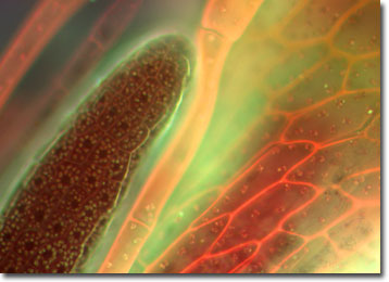
The antheridia and archegonia of mosses are generally found at the tips of the main plant shoots. In some species, the shoots are unisexual, each shoot containing either archegonia or antheridia. Other species have bisexual shoots, containing both archegonia and antheridia.
Like other plants, most mosses reproduce through the alternation of generations, alternating generations of sexual and asexual forms; each complete life cycle requiring two generations. The sexual form, called the gametophyte, begins as a protonema. The protonema is the direct product of spore germination and gives rise to the green leafy plant commonly regarded as a moss. The asexual form, or sporophyte, develops as separate male and female plants, but neither can exist independently. Each sporophyte plant is composed of a capsule, which is the center of spore formation; a stalk; and a foot that attaches the sporophyte body to the tip of the gametophyte. Eventually, the diploid spores are released and, upon successful germination, grow into another moss plant.
The specimen presented here was imaged with a Nikon Eclipse E600 microscope operating with fluorite and/or apochromatic objectives and vertical illuminator equipped with a mercury arc lamp. Specimens were illuminated through Nikon dichromatic filter blocks containing interference filters and a dichroic mirror and imaged with standard epi-fluorescence techniques. Specific filters for the moss archegonia stained thin section were a UV-2E/C, B-2E/C, and a Y-2E/C. Photomicrographs were captured with an Optronics MagnaFire digital camera system coupled to the microscope with a lens-free C-mount adapter.
BACK TO THE FLUORESCENCE DIGITAL IMAGE GALLERY