
Fluorescence Digital Image Gallery
Human Spinal Cord
The human nervous system carries stimuli from sensory receptors to the brain by two main parts: the central nervous system and the peripheral nervous system. The brain and spinal cord make up the central nervous system. The spinal cord represents the major nerve track in all vertebrates.
In humans, the spinal cord extends from the base of the brain to the middle of the back and is about 18 inches long. It is composed of 31 paired spinal nerves that transmit information about internal and external conditions to the brain, and also distribute impulses that mediate reflex actions from the brain to other parts of the body. Essentially, all thoughts and actions depend on the proper functioning of the nervous system. Any disturbance in the nervous system can affect the entire body.
Spinal cord injury (SCI) refers to any injury of the nerves within the spinal canal. The effect of SCI depends on the level and type of the injury, but can result in the loss of mobility and feeling in the body. The type of injury can be broken into two classifications: complete and incomplete. Complete injury results in 100 percent loss of function below the level of the injury site. Incomplete injury, however, results in only some loss of function below the level of injury. The level is determined by the location along the spine at which the injury occurs. The closer the injury is to the brain, the greater the loss of sensation and movement.
Traumas, including car accidents, shootings, and sports related accidents, frequently cause spinal damage. Diseases, such as polio, and birth defects, such as spina bifida, can also result in spinal injury. Over 400,000 people in the United States suffer with SCI and the majority, 82 percent, are men between the ages of 16 and 30. Many are paraplegics, which are unable to move or feel their lower body. Others are quadriplegic, meaning that they have lost feeling and movement in both their upper and lower body.
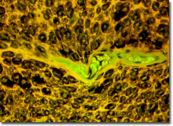
The specimen presented here was imaged with a Nikon Eclipse E600 microscope operating with fluorite and/or apochromatic objectives and vertical illuminator equipped with a mercury arc lamp. Specimens were illuminated through Nikon dichromatic filter blocks containing interference filters and a dichroic mirror and imaged with standard epi-fluorescence techniques. Specific filters for the human spinal cord thin section were a B-2E/C and a Y-2E/C. Photomicrographs were captured with an Optronics MagnaFire digital camera system coupled to the microscope with a lens-free C-mount adapter.
BACK TO THE FLUORESCENCE DIGITAL IMAGE GALLERY