
Fluorescence Digital Image Gallery
Transformed African Green Monkey Kidney Fibroblast Cells (COS-7 Line)
The COS-7 cell line was derived by Yakov Gluzman in the early 1980s from the previously established CV-1 African green monkey kidney line by transformation of the normal cells with an origin defective mutant of simian virus 40 (SV40) that codes for the wild-type virus T-antigen. The fibroblast line grows adherently to glass and plastic in culture and is generally utilized as a transfection host.
COS-7 cells are susceptible to SV40 (tsA209 strain) at 40 degrees Celsius, and SV40 mutants with deletions in certain regions. Because of this susceptibility, COS-7 cells have been utilized extensively in research, and the cell line is often an excellent choice for transfection experiments with recombinant plasmids. In addition, SV40 itself is a popular subject of study largely due to the fact that the small DNA tumor virus features a relatively simple genetic organization and a genome that can be manipulated with relative ease. Moreover, SV40 has been of exceptional scientific interest due to its possible association with certain types of human cancers, especially mesothelioma.
Enclosed in a spherical capsid, SV40 DNA is 5243 nucleotides in size and exhibits a closed circular superhelical conformation. The capsid that encloses the supercoiled DNA is so small that there is only enough space for the genome to encode a small number of functions. The lifecycle of the virus is controlled by a regulatory portion of the genome, which also encodes three capsid proteins. Space is so limited in the genome that the capsid proteins are encoded with reading frames that overlap, so that the final nucleotide sequence of the gene for one protein also encodes for the initial portion of another protein. In addition to capsid proteins, SV40 DNA encodes the potentially tumor-causing protein T-antigen and a spliced version of the protein called small t-antigen. In primate cells, T-antigen usually controls SV40 DNA replication as well as the packaging of the virus into capsids, but in many other cells the protein binds to p53 and Rb proteins, which are important in the regulation of growth, essentially blocking their normal processes and resulting in the development of tumors.
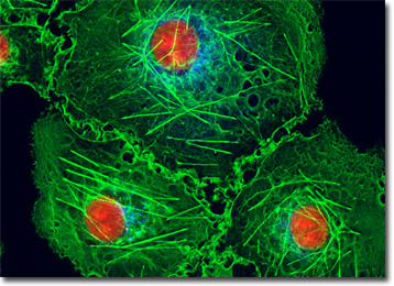
The culture of transformed African green monkey kidney cells featured in the digital image above was labeled with Alexa Fluor 350 conjugated to wheat germ agglutinin, a fluorescent lectin that selectively binds to sialic acid residues, which are found in both mucoproteins and glycoproteins. The cells were also stained with Alexa Fluor 488 conjugated to phalloidin and the intercalating dye propidium iodide, which target the cytoskeletal filamentous actin network and nuclear DNA, respectively. Images were recorded in grayscale with a QImaging Retiga Fast-EXi camera system coupled to an Olympus BX-51 microscope equipped with bandpass emission fluorescence filter optical blocks provided by Omega Optical. During the processing stage, individual image channels were pseudocolored with RGB values corresponding to each of the fluorophore emission spectral profiles.
Additional Fluorescence Images of African Green Monkey Kidney (COS-7) Cells
African Green Monkey Kidney Fibroblasts with Fluorescein, Alexa Fluor 568, and DAPI - An adherent COS-7 culture was immunofluorescently labeled with primary anti-bovine alpha-tubulin mouse monoclonal antibodies followed by goat anti-mouse Fab fragments conjugated to fluorescein. In addition, the cells were simultaneously stained for DNA with the ultraviolet-absorbing probe DAPI, and for the cytoskeletal filamentous actin network with Alexa Fluor 568 conjugated to phalloidin.
Golgi Apparatus in African Green Monkey Kidney Cells - In this section, a culture of transformed African green monkey kidney (COS-7 line) cells is presented that was labeled with Alexa Fluor 488 conjugated to the lectin wheat germ agglutinin. Fluorescent wheat germ agglutinin conjugates are often used as probes for the Golgi network in mammalian cultures. The cells were also stained with Alexa Fluor 568 conjugated to phalloidin and DAPI, which target F-actin and DNA, respectively.
COS-7 Cells with MitoTracker Red CMXRos, Alexa Fluor 488, and Hoechst 33258 - A culture of COS-7 fibroblasts was labeled for mitochondria with MitoTracker Red CMXRos, and for the cytoskeletal filamentous actin network with Alexa Fluor 488 conjugated to phalloidin, a cyclic peptide derived from the toxic death cap fungus (Amanita phalloides). In addition, the fibroblasts were counterstained for DNA in the cell nucleus with Hoechst 33258.
Transformed African Green Monkey Kidney Fibroblast Cellular Microtubular Network - The African green monkey kidney (COS-7) fibroblast cell that appears in this section was resident in a culture that was immunofluorescently labeled with primary anti-tubulin mouse monoclonal antibodies followed by goat anti-mouse Fab fragments conjugated to Cy3, targeting the extensive cellular microtubular network. The culture was also stained for DNA with the ultraviolet-absorbing probe DAPI.
Sialic Acid Residue Distribution in COS-7 Cell Cultures - Similar to the culture of COS-7 cells presented in the digital image above, the African green monkey kidney fibroblasts featured in this section were labeled with Alexa Fluor 350 conjugated to the fluorescent lectin wheat germ agglutinin, which selectively binds to sialic acid residues, primarily in the Golgi apparatus. In addition, the culture was stained for F-actin with Alexa Fluor 488 conjugated to phalloidin, and for nuclear DNA with propidium iodide.
COS-7 Kidney Cells with Cy3, Alexa Fluor 488, and DAPI - An adherent culture of COS-7 African green monkey kidney fibroblast cells was immunofluorescently labeled with primary anti-tubulin mouse monoclonal antibodies followed by goat anti-mouse Fab fragments conjugated to Cy3, targeting microtubules. The cells were simultaneously stained for the cytoskeletal filamentous actin network with Alexa Fluor 488 conjugated to phalloidin, and for DNA with DAPI.
Distribution of Golgi Bodies, F-Actin, and DNA in COS-7 Cells - In this section, the culture of transformed African green monkey kidney cells was labeled with Alexa Fluor 488 conjugated to wheat germ agglutinin, a fluorescent lectin that selectively binds to sialic acid residues, primarily in the Golgi apparatus. The specimen was also labeled with Alexa Fluor 568 conjugated to phalloidin and DAPI, targeting F-actin and DNA in the cell nucleus, respectively.
Transformed African Green Monkey Kidney Fibroblasts with FITC and DAPI - The single cell featured in this section was resident in a COS-7 culture immunofluorescently labeled with primary anti-tubulin mouse monoclonal antibodies followed by goat anti-mouse Fab fragments conjugated to fluorescein isothiocyanate (FITC), which has an absorption maximum of 495 nanometers. The culture was counterstained for DNA in the cell nucleus with the ultraviolet-absorbing probe DAPI.
COS-7 Fibroblast Culture Labeled with Soybean Agglutinin - An adherent culture of COS-7 fibroblasts was labeled with Oregon Green 488 conjugated to soybean agglutinin, a lectin isolated from Glycine max that selectively binds terminal alpha- and beta-N-acetylgalactosamine and galactopyranosyl residues. In addition, the cells were labeled with Alexa Fluor 568 conjugated to phalloidin and DAPI, targeting the cytoskeletal F-actin network and DNA, respectively.
African Green Monkey Kidneys Cells with Alexa Fluor 350, Alexa Fluor 488, and Propidium Iodide - The cytoskeletal F-actin network and nuclear DNA present in a culture of COS-7 cells were targeted with Alexa Fluor 488 conjugated to phalloidin and the intercalating dye propidium iodide, respectively. The specimen was also labeled with Alexa Fluor 350 conjugated to wheat germ agglutinin, a fluorescent lectin that selectively binds to sialic acid residues, which are found in both mucoproteins and glycoproteins.
Microtubule, F-Actin, and DNA Distribution in COS-7 African Green Monkey Kidney Cell Cultures - A culture of transformed African green monkey kidney cells was immunofluorescently labeled with primary anti-tubulin mouse monoclonal antibodies followed by goat anti-mouse Fab fragments conjugated to Cy3, targeting intracellular microtubules. The culture was also stained for DNA with DAPI, and for F-actin with Alexa Fluor 488 conjugated to phalloidin.
Intracellular Mitochondria in Transformed African Green Monkey Kidney Fibroblasts - In the digital image featured in this section, the mitochondria present in a culture of COS-7 kidney fibroblasts are easily observable due to staining with MitoTracker Red CMXRos. The filamentous actin and cell nuclei present in the culture, which were respectively labeled with Alexa Fluor 488 conjugated to phalloidin and Hoechst 33258, are also readily apparent.
BACK TO THE CULTURED CELLS FLUORESCENCE GALLERY
BACK TO THE FLUORESCENCE GALLERY