
Differential Interference Contrast Image Gallery
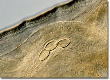
Whipworm (Trichuris) Eggs
Whipworms are small, intestinal parasites belonging to the genus Trichuris that are named for their characteristic tapered shape. More than 60 species of whipworms are capable of infecting mammals, but many exhibit a relatively high level of host specificity.
The sole cause of whipworm infection is ingestion of Trichuris eggs. The eggs, which are expelled by infected individuals with bodily wastes, are quite resilient, however, and may survive in soil or on other surfaces for significant periods of time. When a suitable host ingests an infective egg, it hatches in the small intestine and the juvenile worm migrates to the large intestine. There the parasite embeds its anterior end into the intestinal lining where it reaches sexual maturity and remains for the rest of its lifecycle, which may be several years long.
The whipworm that most often infests humans is Trichuris trichiura. Many of those that are infected with the parasites, however, do not exhibit symptoms. Only when large populations are present in a host do such problems as anemia, pallor, and dysentery occur. Yet, children may suffer more severely from parasite infections than adults, and chronic whipworm infection may lead to mental or physical retardation in the young. Diagnosis of the condition can be difficult, but may easily be treated with medications once the parasitic cause is identified.