
Differential Interference Contrast Image Gallery
Moss Bulbils
Mosses are eukaryotic plants belonging to the division Bryophyta that most often inhabit moist, shady areas. Mosses do not bear seeds or flowers, but they are capable of reproducing in a variety of ways, their lifecycle involving alternate generations of gametophytes and sporophytic plants.
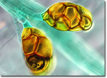
The lush, green mosses that most people associate with wooded areas belong to the dominant gametophyte moss generation. These plants germinate from haploid moss spores that come into contact with incident light. Developing filaments of haploid cells known as protonemata sprout from the walls of the spores in proper conditions and resemble the appearance of filamentous algae. From the tangle of branching protonemata filaments, small, brown bulbs called bulbils may develop. Bud-like, the aerial bulbils develop into typical moss gametophytes when they arrive at a suitable location.
Wide distribution of mosses has played an important part in making Earth inhabitable for more advanced life forms. Mosses have existed for more than 200 million years, appearing sometime after the algae and fungi. The mosses survived in the small amount of nutritive soil that was formed by the decaying material of the more primitive plants, breaking down exposed substrata and releasing nutrients. The remnants of dead mosses then created an even thicker layer of rich soil, making it possible for more complex plants to grow on the surface of the planet.