
Differential Interference Contrast Image Gallery
Moss Antheridia
Reproduction of mosses, an advanced group of the green seedless plants known as Bryophytes, may take many forms. New plants may develop through branching, fragmentation, regeneration, or production of spores.
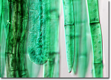
In the gametophyte form of mosses, reproduction is generally sexual and is seasonally controlled. Male sex organs known as antheridia and female sex organs, which are referred to as archegonia, are typically located at the tips of the main shoots of gametophyte mosses. Whether shoots are unisexual or bisexual is a species dependent characteristic, but many mosses are designed to discourage inbreeding. The reproductive process itself can only take pace when the plant is wet, sperm released from antheridia swimming to female archegonia to penetrate ova and create a zygote.
Zygotes remain in the archegonia and undergo several cell divisions that result in the development of embryonic sporophytes. A protective, sheath-like structure called the calyptra encases each embryo. Through intake of water, minerals, and nutrients from the gametophyte, the sporophyte grows in size. When mature, each sporophyte is accompanied by a spore-producing sporangium that sheds its spores when conditions are favorable. In many moss varieties, specialized structures help expel spores away from the parent plant.