
Differential Interference Contrast Image Gallery
Hydatid Cysts
A hydatid is a cyst that houses an intermediate form of the tapeworm Echinococcus granulosus. Hydatids may be found in animals such as sheep, cattle, goats, and humans, but the adult parasites only inhabit dogs and other canine species.
Intermediate hosts become infected with hydatid cysts, a condition known as hydatid disease, by accidentally consuming the eggs of E. granulosus adults. The eggs, once expelled with bodily wastes from infected definitive hosts, may survive for long periods of time even in harsh environments. When a suitable intermediate host ingests them, the eggs hatch in the small intestine and penetrate the wall of the gut to enter the circulatory system. The larvae are then distributed to various locations within the body and grow into hydatid cysts.
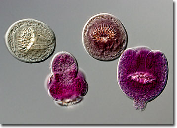
Hydatid cysts are typically spherical in shape and have the ability to achieve rather large sizes. The insides of the cysts are filled with fluid, brood capsules, daughter cysts, and protoscolices that have the capability to grow into adult worms if consumed by a definitive host. If a cyst is ruptured, which may occur through a sharp blow or during surgery, each protoscolex released may form a new cyst. Also, the fluid within the hydatids is highly allergenic and may cause anaphylactic shock and rapid death if freed inside the body.
In most intermediate hosts, hydatid cysts go unnoticed. In humans, however, clinical signs of hydatid disease vary depending on the location of cyst development and the size they achieve. The majority of cysts grow in the tissues of the liver, but may also settle in bone marrow, the peritoneal cavity, or other organs, including the lungs, kidneys, and brain. Hydatid cysts tend to cause pressure related injuries in human hosts and are most serious when they are extremely large or are housed in the vicinity of vital organs.