
Differential Interference Contrast Image Gallery
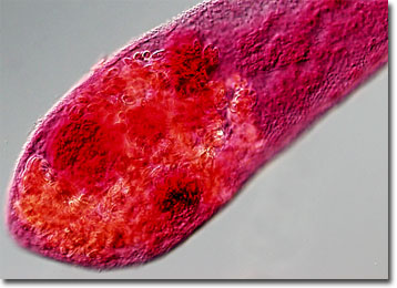
Intestinal Fluke (Heterophyes)
Members of the genus Heterophyes are intestinal flukes that average 1.5 millimeters in length. The most common of the ten species that belong to the genus is Heterophyes heterophyes, which is responsible for the condition known as heterophyiasis in humans, but may also be found in foxes, dogs, and cats.
Humans and other mammals become infected with the digenetic trematode by eating contaminated raw or undercooked fish. Thus, most cases of heterophyiasis occur in the Far East, the Middle East, and Egypt due to the characteristic diets of inhabitants of these areas. True levels of incidence are difficult to discern, however, because cases are often asymptomatic and signs of serious occurrences, such as abdominal pain, diarrhea, and the frequent appearance of eggs in feces, are similar to those of other types of parasite infection. In rare instances, H. heterophyes can cause the lining of the small intestine to break down, and the eggs produced by the parasite enter into the blood stream. Once in the blood stream the eggs may be carried to other organs where they can instigate acute medical problems.
H. heterophyes must inhabit two intermediate hosts before it is capable of infecting a human, or other definitive host. In order to hatch, eggs of the parasite must first be ingested by a suitable snail. Within the snail, larvae penetrate its tissues and develop through two morphologically distinct generations. The motile larvae that result exit the mollusk host and seek out suitable fish, which they invade to form their mammalian infective stage. Once the infected fish are eaten by a definitive host, they make their final home in the small intestine, developing into sexually mature adults and producing a new generation of embryonated eggs.