
Differential Interference Contrast Image Gallery
Trematode (Fasciolopsis buski) Rediae
A parasitic trematode, Fasciolopsis buski is known as the giant intestinal fluke of man, but may also infect swine. In their adult form, the worms attach themselves to the tissues of the small intestine through the use of ventral suckers and may reach up to 7.5 centimeters in length.
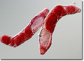
F. buski was first described in 1843 by English surgeon George Busk, who found the parasite in the duodenum of a sailor. The complex lifecycle of the trematode was not understood, however, until Claude H. Barlow’s determination of it in 1925. As he revealed, immature eggs of F. buski are discharged from humans or other definitive hosts with bodily wastes. If the eggs then reach water, they hatch in 3 to 7 weeks and form miracidia, organisms that looks similar to ciliated protozoa. The miracidia penetrate the soft tissues of snails and develop into a sporocyst state. The sporocysts then produce many daughter stages called rediae that asexually reproduce to yield cercariae. The cercariae, which feature a tail-like structure that enables them to swim, exit the snail host and encyst on nearby vegetation, where they develop into metacercariae.
Humans become infected with F. buski, a condition known as Fasciolopsiasis, by consuming uncooked aquatic vegetables, such as lotus, water chestnuts, and water bamboo, contaminated with metacercariae. Generally settling in the duodenum, the metacercariae become adult worms in approximately 3 months. Though infection is sometimes asymptomatic, at the site of attachment deep inflammatory ulcerations often occur, usually accompanied by nausea, abdominal pain, and diarrhea, alternated with periods of constipation. Particularly large populations of F. buski can also involve mucus discharge, general body weakness, and fluid retention that may result in severe consequences if left untreated.