
Differential Interference Contrast Image Gallery
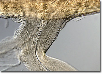
Earthworm Nervous Tissue
School children are often squeamish the first time they are required to dissect an earthworm for class. Advanced research carried out with the creatures, however, has proved invaluable to the scientific world, and recent studies of the segmented worms may provide clues of how to quickly repair severed nerves.
Earthworms do not have eyes or ears, but are perceptive of light and sound. This strange phenomenon is possible because the earthworm nervous system is connected to a variety of sensory cells that are capable of detecting different environmental factors. Earthworms also have a developed sense of touch, a sense of taste, and can perceive the amount of moisture in surrounding soil. Some studies have even shown that earthworms can store simple memories, such as which branch of a Y-shaped tube should be taken to avoid an electrical shock.
The nervous system of an earthworm is composed of a primitive brain of fused ganglia, a ventral nerve cord, and peripheral nerves. Impulses from the earthworm’s sensory cells are transmitted by the peripheral nerves to certain parts of the body and proper responsive movements are coordinated. If sensory cells indicate that the area outside its burrow is too hot or too cold, for instance, the earthworm will readjust its position so that its less vital, posterior portion is facing that direction. When the organism senses that conditions outside are favorable, it resides with its head towards the surface.