
Differential Interference Contrast Image Gallery
Ctenoid Fish Scale
Ctenoid scales are common to most varieties of bony fishes and feature a comb-like, spiny posterior edge. Their shape is the source of their name, which was derived from the Greek word cteno, meaning “comb.”
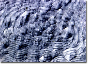
Three general types of ctenoid scales have been categorized, but each type is composed of two key sections; a rigid surface layer chiefly consisting of crystallized calcium salts and a deeper layer that is formed by collagen-type fibrils. The distinction between the ctenoid scale types, therefore, is primarily based upon their form. Crenate scales, for instance, feature simple indentations on their edges, while spinoid scales are characterized by a cluster of spines that extend from the main scale body. Basic ctenoid scales, on the other hand, exhibit ctenii, which are formed as separate bony growths distinct from the main body of the scale.
Similar to trees, the age of a fish may be estimated by examining growth rings on its ctenoid scales. This is because as a fish grows, so does its scales. The increase in size leaves rings, known as circuli, which are similar to those that appear in tree trunks. In winter months or other periods of cold weather, the growth rate of fish and their scales decelerates. Consequently, the circuli occur at much smaller intervals, which leaves a dark band. By counting the bands, known as annuli, an approximate age of a fish may be obtained.