
Wenham's Universal
Inclining and Rotating
Microscope
The London scientific instrument firm started by Andrew Ross in the early 1830s produced a number of remarkable microscopes including this beautiful specimen in 1882. Although the microscope was signed "Ross London", it was actually designed and built by Francis Wenham.
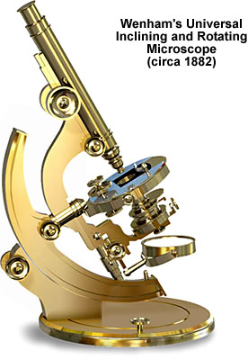
This microscope is part of the Royal Microscopical Society collection and has been described in detail by Gerard Turner in his comprehensive and informative treatise The Great Age of the Microscope. Designed to feature a wide range of latitude in the observation of both transmitted and obliquely-illuminated samples, the microscope is almost infinitely-configurable. The body tube assembly rests on a long curved solid-brass limb that is attached to a flat circular base. Both the limb and base have a great freedom of movement, with the limb being capable of large inclinations to either side of the microscope axis and the base can be rotated 360-degrees. A circular stage, also capable of complete rotation, is attached to a smaller limb containing the body tube, which can also be inclined. Various models of this microscope contain either a monocular body tube or a Wenham binocular tube. The rim of the circular base is graduated in 1-degree marks through 360 degrees and the stage is also graduated in 0.5-degree increments. Objectives and oculars were of high quality compared to other optics of the period.
BACK TO NINETEENTH CENTURY MICROSCOPES