
Jan Swammerdam Single Lens Microscope
Jan Swammerdam was a seventeenth century Dutch microscopist who favored single-lens microscopes. The microscope featured below was employed by Swammerdam sometime in the mid-1670s and utilizes small bead lenses (one or two millimeters in diameter) that produce a magnification of around 150x, similar to the microscopes of Antoni van Leeuwenhoek.
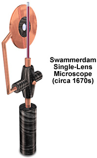
In March of 1678, Swammerdam included drawings of the microscope illustrated above in a letter to his mentor that described several experiments and observations. The single-lens microscope bears a striking resemblance to instruments made during this period by Johan Joosten van Musschenbroek in Leiden. Effectively a very small magnifying glass, the microscope is designed to be held in one hand while observing specimens. In practice microscopes having this design are very difficult to use because the specimen almost touches the lens, while the observer has to place their eye close to the lens in order to view the specimen. Typically, it is very difficult to discern much of the specimen detail. Swammerdam warned the readers of his Book of Nature that the lens:
"must, for this purpose, be carefully managed, for as it is turned one way or another, different things are seen; one cannot bring the lens nearer, or remove it further, by the least distance, but something is immediately perceived by the sight, which was not observed before."
In his book, Swammerdam indicated that he only observed specimens visible under direct natural light, generally outdoors on summer mornings. In several of his letters he wrote that observations were halted in the autumn and winter due to lack of sufficient light. Prior to his microscopic observation of specimens, Swammerdam carried on painstaking dissections with a variety of tools including fine pairs of scissors, a saw made from a small section of watch spring, a fine sharp-pointed pen-knife, feathers, glass tubes, small tweezers, needles and forceps. He utilized a variety of original and highly effective techniques to clean the specimen and to dissolve unwanted tissues and highlight those of interest. Without a camera to capture images, Swammerdam made drawings of his specimens, first in red crayon, then completed in black ink or pencil. Many of the drawings were ultimately transferred to copper plates for printing.
The microscope illustrated above is accompanied by a specimen holder designed to examining blood samples. The glass tube is filled with blood, which is then observed through the small bead lens mounted in ebony. A small flexible copper clasp is used to position the lens assembly with respect to the specimen for focusing. The instrument sports an ebony handle that is used to position the microscope and specimen near the observer's eye.
Contributing Authors
Matthew Cobb - Laboratoire d'Ecologie, CNRS-Université, Paris 6, Paris, France.
Michael W. Davidson - National High Magnetic Field Laboratory, 1800 East Paul Dirac Dr., The Florida State University, Tallahassee, Florida, 32310.
BACK TO SIXTEENTH-SEVENTEENTH CENTURY MICROSCOPES