
French Compound Monocular Microscope
A ball-and-socket joint enables easy adjustment of the inclination angle on this French compound microscope, which was constructed around 1875. The illustration of the brass monocular instrument presented here is based on a photograph and description of the original model, which is part of the Billings Collection of the Walter Reed Army Hospital in Washington DC.
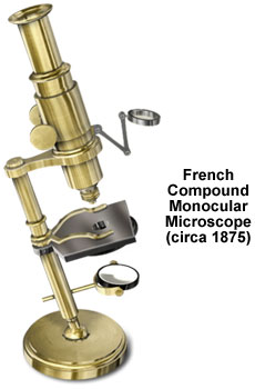
The circular, bronze base of the microscope supports a 6-inch tubular pillar mounted on the ball-and-socket joint. Attached to the lower portion of the pillar is a gimbal-mounted, 1.2-inch diameter mirror and above the stage, a condenser of the same size is suspended by a jointed arm. The fixed, rectangular stage contains a circular aperture and a revolving disc of diaphragms attached to its underside. A U-shaped slide holder on sliding casing mounts is also included to secure specimens to the upper surface of the stage.
At the top of the cylindrical brass pillar, a screw holds the 2.5-inch arm and its ring to the microscope stand. The fixed tube of the instrument features a single, milled-head pinion, while the 6.25-inch body tube carries the rack for the coarse focusing mechanism. Fine focus is not provided on this basic French microscope, but its optical system includes a Huygenian eyepiece, a single objective, and an internal field lens. Both Nachet & Son and E. Hartnack & Company made similar unsigned trade microscopes in their Paris workshops.
BACK TO NINETEENTH CENTURY MICROSCOPES