
George Adams Jr. Lucernal Microscope
Although not signed, the lucernal microscope portrayed here is almost certainly the work of British instrument maker and optician George Adams Jr., who would have designed it around 1790. A photograph and description of the original wooden and brass optical instrument is featured in Gerard Turner's book entitled The Great Age of the Microscope.
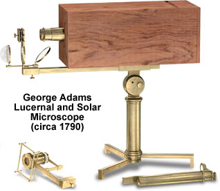
In lucernal microscopes, the observer's eye and the objective are conjugate with a large convex lens mounted on one end of a body tube. Light from the objective is projected from the condensing lens onto the eye or a ground glass focusing screen. In the illustration above, however, an end piece of the rectangular mahogany housing covers the focusing screen, a feature that provides protection from damage and dirt when the microscope is not in use. Also, the tripod foot that supports the brass cylindrical pillar (topped by a compass joint) may be folded flat to reduce storage space requirements.
Focus of this instrument is accomplished by turning either of the knobs that are mounted on the pillar, an action that moves the stout, horizontal brass bar supporting the microscope back and forth. A stage at the front of the horizontal bar, which is designed for holding opaque specimens, is illuminated by a bull's-eye condenser and a concave mirror. The compound, 205-millimeter body tube of the instrument is fitted with an eyepiece composed of two biconvex lenses and a biconvex field lens. A brass-mounted projection lens that protrudes from the wooden projection box completes the device's optical system.
Accompanying the instrument is a brass, telescoping simple microscope, featured in the lower right-hand corner of the illustration. The smaller microscope serves as an eye-locator for high-powered objectives that do not adequately illuminate the focusing screen. There is also a spring stage in the lower left-hand corner that features a condenser in a sliding tube and rectangular frame, which can be used to tie down frogs and other specimens for observation or dissection.
BACK TO EIGHTEENTH CENTURY MICROSCOPES