
Interactive Java Tutorials
The Critical Angle of Reflection
An important concept in optical microscopy is the critical angle of reflection, which is a necessary factor to consider when choosing whether to use dry or oil immersion objectives to view a specimen at high magnification. Upon passing through a medium of higher refractive index into a medium of lower refractive index, the path taken by light waves is determined by the incident angle with respect to the boundary between the two media. This interactive tutorial explores the transition from refraction to total internal reflection as the angle of the incident wave is increased at constant refractive index.
The tutorial initializes with an incident light wave (represented by a sine function) emerging from a water-air interface at an incident angle of 30 degrees. At the interface, the light wave is refracted by the angle q(r) and passes through the air in a straight trajectory after being deviated. In order to operate the tutorial, use the Incident Angle slider to adjust the value of the incident light wave (impacting the interface) between a value of zero and 60 degrees. At the critical angle for the material through which the incident light ray propagates (48.75 degrees for the default material, water), the light will be totally internally reflected at the interface back into the material without passing through and undergoing refraction. The Wavelength slider can be employed to vary the wavelength of the incident, refracted, and reflected waves. After a specific medium has been examined, a new material having a different refractive index (and critical angle) can be selected from the Choose A Material pull-down menu. The refraction or reflection angle at the interface is continuously calculated using Snell's Law, and displayed beneath the sliders.
If the incident angle increases past a specific value (dependent upon the refractive index of the two media), it reaches a point at which the angle is so large that no light is refracted into the medium of lower refractive index, as illustrated in Figure 1. In this figure, individual light rays are represented by either red or yellow colored arrows moving from a medium of higher refractive index (n(2)) to one of lower refractive index (n(1)). The angle of incidence for each individual light ray is denoted by the value i and the angle of refraction by the variable r. The four yellow light rays all have an angle of incidence (i) low enough to allow them to pass through the interface between the two media. However, the two red light rays have incident angles that exceed the critical angle of reflection (approximately 41 degrees for the water and air examples) and are reflected either into the boundary between the media or back into the higher refractive index medium.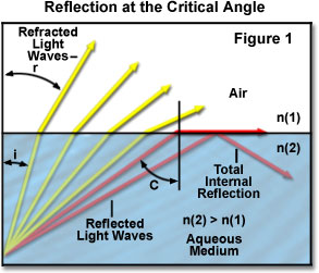
The critical angle phenomenon takes place when the angle of refraction (angle r in Figure 1) becomes equal to 90 degrees and Snell's law reduces to:
where (q) is now termed the critical angle c. If the medium having a lesser refractive index is air (n = 1.00), the equation reduces to:
When the critical angle is exceeded for a particular light wave, it exhibits total internal reflection back into the medium. Usually the higher index medium is considered the internal medium, because air (having a refractive index of 1.0) is in most cases the surrounding, or external medium. This concept is especially critical in optical microscopy when attempting to image specimens with a medium other than air between the cover glass and the objective front lens. The most common immersion medium (other than air) is specialized oil having a refractive index equal to that of the glass used for the objective front lens element and the coverslip.
Contributing Authors
Robert T. Sutter, Thomas J. Fellers, and Michael W. Davidson - National High Magnetic Field Laboratory, 1800 East Paul Dirac Dr., The Florida State University, Tallahassee, Florida, 32310.