
Image Formation
Interactive Java Tutorials
Axial Resolution and Depth of Field
The lateral resolution for an Airy diffraction pattern generated by a point light source is defined within a single plane of focus at the intermediate image position in an optical microscope. In fact, the diffraction image of a point source extends periodically and symmetrically above and below this plane into a three-dimensional pattern that expands and spreads out from the center along the optical axis. This tutorial explores the structure of cross sections taken along the optical axis of the microscope near the focal plane using a virtual high numerical aperture objective free from spherical aberration.
The first image displayed by the tutorial represents an oblique view of a longitudinally sectioned three-dimensional diffraction pattern that extends along the optical axis of the microscope, near the point of paraxial focus (the intermediate image plane). As the objective lens is focused a very short distance above and below the point of exact focus, a two-dimensional Airy diffraction pattern of the point illumination source is formed in the intermediate image plane. Pattern intensity oscillates between bright and dark regions as cross sections are taken through the diffraction pattern at different focal points above and below the point of paraxial focus. As the objective is focused increasing farther away from the paraxial focal point, the outer diameters of the Airy patterns expand, in some cases with a reversal of contrast in the central region, and the absolute intensity diminishes.
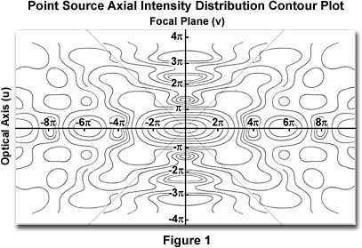
To operate the tutorial, use the blue buttons to toggle between focal planes surrounding the point of paraxial focus along the optical axis of the microscope. It is helpful to refer to the isophot of a longitudinal section taken from the three-dimensional diffraction image presented in Figure 1. In this figure, the microscope optical axis is represented by the abscissa (u) and extends for a distance of ± 10p, while the intermediate image (focal) plane is located by the ordinate (v) and covers a range of ± 4p. The first cross section presented in the tutorial occurs at 6p along the optical axis, and illustrates the an expanded region of the diffraction pattern having a central circular maximum surrounded by higher-order diffraction rings. As successive cross sections that lie increasingly closer to the intermediate image plane are examined (in 2p increments), the three-dimensional diffraction pattern spread decreases and becomes more intensified. A reversal of contrast in the central spot occurs at ±4p, with the spread reverting to a classical Airy pattern having a diffuse central region surrounded by diffraction rings at ±2p. At the intermediate image plane (u=0), which is the point of exact (or paraxial) focus, we observe the classical Airy pattern with a minimum at v=1.22p.
Contributing Authors
Kenneth R. Spring - Scientific Consultant, Lusby, Maryland, 20657.
Matthew J. Parry-Hill and Michael W. Davidson - National High Magnetic Field Laboratory, 1800 East Paul Dirac Dr., The Florida State University, Tallahassee, Florida, 32310.