
Interactive Java Tutorials
Acousto-Optic Tunable Filters
Wavelength selection is of fundamental importance in many arenas of the optical sciences, including fluorescence spectroscopy and microscopy. Electro-optic devices, such as the acousto-optic tunable filter (AOTF), are increasingly being employed to modulate the wavelength and amplitude of illuminating laser light in the latest generation of confocal microscopes. These filters do not suffer from the mechanical constraints, speed limitations, image shift, and vibration associated with rotating filter wheels, and can easily accommodate several laser systems tuned to different output wavelengths. In addition, acousto-optic filters do not deteriorate when exposed to heat and intense light as do fluorescence interference filters.
The tutorial initializes with an Incident Beam of non-polarized light (represented by orthogonal yellow sine waves) impacting the tellurium dioxide crystal of an acousto-optic tunable filter from the left-hand side of the window. The dioxide crystal is sandwiched between an acoustic transducer and absorber that can be regulated by the Acoustic Power (default value of 1.7 watts) and Acoustic Frequency (default value of 250 megahertz) sliders. Upon encountering the standing wave in the tellurium dioxide crystal, a portion of the incident light beam is diffracted into the Confocal Scan Head while the remainder of the beam passes through the crystal and is absorbed by a Beam Stop. In order to operate the tutorial, use the Acoustic Power slider to vary the input power to the standing acoustic wave between a value of 0.5 and 3.0 watts. As the slider is moved to the right or left, the amplitudes of the waves passing through the AOTF are increased or decreased. Wavelength selection is accomplished with the Acoustic Frequency slider, which has a range of 150 to 350 megahertz and can cover the entire spectral region between 400 and 700 nanometers.
In an acousto-optic tunable filter, a piezoelectric transducer bonded to a crystal of tellurium dioxide or quartz generates high-frequency acoustical compression waves that alter the refractive index of the crystal in a periodic pattern. This leads, in effect, to the generation of a diffraction grating so that an orthogonal beam of polarized and collimated light incident at the Bragg angle is diffracted with high efficiency into the first order beam (the diffracted beam in the tutorial). Variations in the frequency of the acoustic waves alter the spacing pattern in the crystal and, hence, the wavelength of the diffracted light (the Acoustic Frequency slider). Likewise, altering the amplitude of the acoustic wave (with the Acoustic Power slider) determines its relative intensity. The diffraction efficiency can reach levels of 85 percent and the switching speed for both wavelength and power is in the megahertz range. A typical AOTF can accept incident light having a maximum half-cone angle of approximately 5 degrees, restricting the use of these devices to well-collimated light sources.
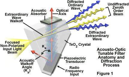
Acousto-optic filters are configured to operate through either a collinear (for example, using quartz as the acoustical crystal) or a noncollinear mechanism, depending upon device design parameters. In a collinear AOTF, incident light and the standing acoustic wave interact collinearly within the birefringent crystal to produce a coupled diffracted wavefront that is also collinear to the input beam (and acoustic wave). Because the polarization of the incident light beam is orthogonal to that of the diffracted beam and the two beams are collinear, a polarizer must be used to separate the beams. Conversely, in a noncollinear AOTF (using a tellurium dioxide crystal), the acoustic and optical waves propagate through the crystal at different angles so that the incident (zeroth order) and diffracted beams are physically separated and the filter does not require polarizers. In addition, the two orthogonal wavefronts do not separate until they exit the crystal (see Figure 1), so the diffraction angle (and spatial location of the resulting image) does not change with wavelength.
Presented in Figure 1 is a typical noncollinear AOTF based on a tellurium dioxide crystal, where the acoustic and optical waves propagate at different angles through the crystal. If the input light beam is non-polarized (illustrated in the figure as a focused light cone), the diffracted wavefronts are directed into physically separated first-order beams that are orthogonally polarized (the ordinary and extraordinary diffracted waves). If linearly polarized light is incident on the crystal, only a single diffracted beam having its polarization perpendicular to the incident light is created. In order to use a noncollinear AOTF as a tunable filter, the undiffracted (zeroth order) beam is blocked (as in the tutorial), allowing only the diffracted monochromatic wavefront to enter the confocal scan head and subsequently illuminate the specimen. The angle between the zeroth order and first-order diffracted beams is a function of the AOTF design, but usually ranges over just a few degrees.
In optical microscopy, acousto-optic tunable filters are increasingly being employed for duty with a variety of laser sources as input illumination in confocal scanning heads. However, these devices are also used with laser illumination systems on widefield microscopes, as well as with conventional light sources, such as arc-discharge (mercury and xenon) lamps. By combining the AOTF with a digital signal processor (DSP), up to six different wavelengths can be diffracted simultaneously, each at an individually controlled power level.
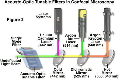
A practical light source configuration scheme utilizing an acousto-optic tunable filter for confocal microscopy is illustrated in Figure 2. The output of three laser systems (helium-cadmium, argon, and argon-krypton) are combined by dichromatic mirrors and directed through the AOTF, where the first-order diffracted beam (purple) is collinear and is launched into a single-mode fiber. The undiffracted laser beams (green, yellow, and red) exit the AOTF at varying angles and are absorbed by a beam stop (not illustrated). The major lines (wavelengths) produced by each laser are indicated (in nanometers) beneath the hot and cold mirrors. The dichromatic mirror reflects wavelengths lower than 525 nanometers and transmits longer wavelengths. Two longer wavelength lines produced by the argon-krypton laser (568 and 648 nanometers) are reflected by the hot mirror, while the output of the argon laser (458, 476, 488, and 514 nanometers) is reflected by the dichromatic mirror and combined with the transmitted light from the argon-krypton laser. Output from the helium-cadmium laser (442 nanometers) is reflected by the cold mirror and combined with the longer wavelengths from the other two lasers, which are transmitted through the mirror.
Using acousto-optic tunable filters in laser scanning confocal microscopy enables the operator to vary the wavelength or intensity of illumination on a pixel-by-pixel basis while maintaining a high scan rate. In this manner, specifically defined regions of interest can be illuminated more intensely for studies employing photobleaching (for example, in fluorescence recovery after photobleaching, FRAP), or at two different wavelengths for spectroscopy and excitation ratio measurements. The geometry of the scanned regions can be arbitrarily defined by selection in an overlay plane so that only features of interest are illuminated under control of the digital signal processor. The size of the selected region is determined by the magnification and numerical aperture of the microscope objective, but the spectral content and intensity of the illumination are under the control of the AOTF device.
Several significant limitations must be addressed when using acousto-optic tunable filters in confocal scanning instruments. The diffraction efficiency of tellurium dioxide-based devices drops markedly at wavelengths below 450 nanometers, restricting applications involving shorter wavelengths (330 to 450 nanometers) to devices based on quartz crystals (which require far more power to operate). In addition, even highly collinear AOTFs exhibit some dispersion that can yield slight beam displacement at the extremes of their wavelength range. This is not usually a problem in confocal microscopy because the tunable filter is in very close proximity to a single-mode fused silica optical fiber equipped with an integral lens system. As a result, AOTF beam shift becomes negligible during the short journey between the filter output and fiber input. Finally, thermal effects resulting from varying acoustic power and frequency combinations can transiently alter the refractive index of the crystal and reduce diffraction efficiency until temperature stability is again achieved. This index shift often produces significant drift in output intensity, which is a serious problem when quantitative analysis of specimen features is based on the assumption of stable and reproducible illumination beam intensity. Acoustically generated thermal effects can be reduced to an insignificant level by heating the device above ambient temperature.
One of the most interesting marriages between acousto-optic filters and confocal microscopy revolves around the simultaneous use of multiple fluorescent probes in living cells, which is an increasingly popular technique for monitoring the assembly of macromolecular complexes, protein-protein interactions, and the targeting of proteins to specific sites in the cell. The ability to label a protein of interest with a fluorophore in a living cell was revolutionized by the development of techniques to incorporate the green fluorescent protein (GFP) and a multiple of relatives into the intracellular protein synthetic machinery. In these experiments, the accumulation of a multispectral image set is critical before movements alter the relationships or geometry of the target specimen features. Acousto-optic tunable filters permit the investigator to control the excitation wavelength and intensity of the probing beam so that the same region can be simultaneously or sequentially illuminated at different wavelengths before any movements occur.
Contributing Authors
Kenneth R. Spring - Scientific Consultant, Lusby, Maryland, 20657.
Matthew J. Parry-Hill, Ian D. Johnson, and Michael W. Davidson - National High Magnetic Field Laboratory, 1800 East Paul Dirac Dr., The Florida State University, Tallahassee, Florida, 32310.