
Optical Aberrations
Interactive Tutorials
Geometrical Distortion
Distortion is an aberration commonly seen in stereoscopic microscopy, and is manifested by changes in the shape of an image rather than the sharpness or color spectrum. The two most prevalent types of distortion, positive and negative (often termed pincushion and barrel, respectively), can often be present in very sharp images that are otherwise corrected for spherical, chromatic, comatic, and astigmatic aberrations. In this case, the true geometry of an object is no longer maintained in the image.
The tutorial initializes with a randomly selected photomicrograph or digital image appearing in the microscope viewport. To select a new image, use the Choose A Specimen pull-down menu. The Distortion slider is utilized to introduce either pincushion (slider to the left) or barrel (slider to the right) distortion into both the image and a horizontal/vertical grid pattern positioned above the slider. As the grid pattern changes to illustrate varying degrees of aberration, the approximate percentage of distortion is displayed beneath the pattern.
Introduction of distortion aberration is more obvious in specimens having a network of regular periodic features, such as diatoms or microprocessors. Other specimens lacking such periodicity do not appear to be dramatically distorted when viewed in the microscope, as evidenced by the biological specimens selectable with the pull-down menu. This does not mean that specimens with irregular features are immune to distortion aberration, just that the aberration must be far more severe to produce noticeable effects.
Although modern research-grade microscopes are corrected in order to keep aberrations from spreading the image of a point source beyond the Airy disk, geometrical distortion of the image formed by microscope objectives tends not to be as well corrected when compared to photographic lenses at the same image angle. Distortion is produced mainly by the oculars (between 5 and 10 percent of the radial distance) in optical microscopy, although some distortion is also found in lower quality objectives. Microscopes can be monitored for distortion by imaging crossed grating lines, such as those found in haemocytometers, in the wide-field mode. When viewed through the eyepieces, the lines should appear straight and parallel over the entire image field.
Most objectives designed for use in biological microscopes can have pincushion distortions up to one percent, however objectives designed for imaging semiconductors are essentially distortion-free. This is necessary because the majority of integrated circuits have surface features with an abundance of grid-like structures composed of horizontal and vertical lines. Any distortion present in objectives when imaging these samples will be obvious in the eyepieces and in photomicrographs. Images produced by objectives, eyepieces and other optical components that have been corrected for geometrical distortion aberration are referred to as orthoscopic images.
The origin of geometrical distortion lies in a difference between the transverse magnification of a lens and the off-axis image distance. When this distance deviates from that predicted by paraxial theory for constant transverse magnification, distortion can arise due to differences in focal lengths and magnifications through various parts of the lens. In the absence of other aberrations, geometric distortion is manifested by a mis-shaped image, even though each image point is in sharp focus, as discussed above. Quantitatively, distortion can be described by the following equation:
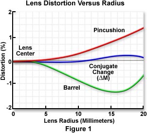
where M is the axial lateral magnification and M(l) is the off-axis magnification at the image plane. If the lateral magnification increases proportionally with the off-axis distance of the object, distortion is positive, producing a pincushion effect (Figure 1). In this instance, each image point is displaced radially outward from the center, with the peripheral image points being translocated the greatest distance. Alternatively, when magnification is decreased with the off-axis object distance, distortion is negative and a barrel aberration is observed. Barrel distortion corresponds to a situation where the transverse magnification decreases with axial distance and each image point moves radially towards the center of the image.
In general, thin lenses show little or no distortion, whereas thicker positive and negative simple lenses will suffer positive and negative distortion, respectively. The sign (or type) and magnitude of geometric distortion depend on the position of the aperture diaphragm with respect to the lens(es). When an aperture diaphragm is placed in front of a positive lens, the principal ray does not form an image at the predicted Gaussian point and barrel distortion occurs, whereas when the diaphragm is placed behind the lens (forming an exit pupil), a pincushion distortion effect is observed.
Distortion is often found in systems utilizing compound lens systems containing meniscus, double gauss, telephoto, retrofocus, fisheye, and zoom lenses. In telephoto and retrofocus lens designs, the front group acts as an aperture stop for the rear group, producing a pincushion distortion for the negative rear group in telephoto lenses and a barrel distortion for the positive rear group in retrofocus lenses. Complex lens systems such as the zoom design can have rather pronounced distortion, which may vary with focal length, producing pincushion distortion at long focal lengths and barrel distortion at short focal lengths. For this reason, stereoscopic zoom microscopes classically have a significant amount of distortion present and microscope manufacturers have expended considerable effort in alleviating this aberration.
Contributing Authors
H. Ernst Keller - Carl Zeiss Inc., One Zeiss Dr., Thornwood, NY, 10594.
Kenneth R. Spring - Scientific Consultant, Lusby, Maryland, 20657.
Matthew J. Parry-Hill and Michael W. Davidson - National High Magnetic Field Laboratory, 1800 East Paul Dirac Dr., The Florida State University, Tallahassee, Florida, 32310.
BACK TO LENSES AND GEOMETRICAL OPTICS