
Depth of Field
and
Image Depth
When considering resolution in optical microscopy, a majority of the emphasis is placed on point-to-point resolution in the plane perpendicular to the optical axis. Another important aspect to resolution is the axial resolving power of an objective, which is measured parallel to the optical axis and is most often referred to as depth of field. Axial resolution, like horizontal resolution, is determined by the numerical aperture of the objective only, with the eyepiece merely magnifying the details resolved and projected into the intermediate image plane.

Just as in classical photography, depth of field is determined by the distance from the nearest object plane in focus to that of the farthest plane also simultaneously in focus. In microscopy depth of field is very short and usually measured in terms of microns. The term depth of focus, which refers to image space, is often used interchangeably with depth of field, which refers to object space.
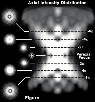
This interchange of terms can lead to confusion, especially when the terms are both used specifically in terms of depth of field. The geometric image plane might be expected to represent an infinitely thin section of the specimen, but even in the absence of aberrations, each image point is spread into a diffraction figure that extends above and below this plane. The Airy disk, discussed in the section on Image Formation, represents a section through the center of the image plane. This increases the effective in-focus depth of the Z-axis Airy disk intensity profile that passes through slightly different specimen planes.
Depth of Field and Image Depth
|
||||||||||||||||||||||||||||||
Table 1
Depth of focus varies with numerical aperture and magnification of the objective, and under some conditions, high numerical aperture systems (usually with higher magnification power) have deeper focus depths than do those systems of low numerical aperture, even though the depth of field is less. This is particularly important in photomicrography because the film emulsion or digital camera sensor must be exposed at a plane that is in focus. Small errors made to focus at high magnification are not as critical as those made with very low magnification objectives. Table 1 presents calculated variations in the depth of field and image depth in the intermediate image plane in a series of objectives with increasing numerical aperture and magnification.
Depth of field equations:
Equation by Charles P. Shillaber from Photomicrography in Theory and Practice on page 254:
Where d represents the depth of field, l is the wavelength of illuminating light, n is the refractive index of the medium (usually air (1.000) or immersion oil (1.515)) between the coverslip and the objective front lens element, and NA equals the objective numerical aperture. Using this equation, depth of field (d) and wavelength (l) must be expressed in similar units. For example, if d is to be calculated in micrometers, l must also be formulated in micrometers (700 nanometer red light is entered into the equation as 0.7 micrometers).
Contributing Authors
Mortimer Abramowitz - Olympus America, Inc., Two Corporate Center Drive., Melville, New York, 11747.
Michael W. Davidson - National High Magnetic Field Laboratory, 1800 East Paul Dirac Dr., The Florida State University, Tallahassee, Florida, 32310.
BACK TO ANATOMY OF THE MICROSCOPE