
Brightfield Microscopy Digital Image Gallery
Prostate Gland (Older)
The prostate gland is an important part of the male reproductive system that contributes to the seminal fluids that contain sperm. Positioned directly below the bladder, the gland surrounds the upper part of the urethra, the duct responsible for emptying urine from the bladder.
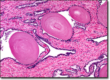
View an image of the older prostate gland at 20x magnification.
Following puberty, the prostate gland is about the size of a walnut for much of a man’s life. However, around the age of 40 or 50, the prostate often begins to enlarge, a process referred to as benign prostatic hypertrophy (BPH). The reason for this physiological change is not completely understood, but is believed to involve a number of factors, such as increased levels of the female sex hormone estradiol and greater production of dihydrotestosterone, a derivative of the male sex hormone testosterone. When the prostate enlarges to a significant extent, symptoms of the condition, which chiefly consist of urination problems caused by the pressure of the gland on the urethra, usually ensue.
The number of men that suffer from an enlargement of the prostate gland later in life is on the rise. Fortunately, in many cases this growth is non-cancerous. However, as instances of BPH increase, so do the number of sufferers of prostate cancer, one of the most common of all types of cancer. Indeed, it is already generally believed that most men have at least a small cancerous nodule in the prostate gland by the time they are 80, although many never realize it. In some cases, however, prostate cancer may spread to other areas of the body and cause serious problems. Thus, regular prostate exams are recommended, and younger individuals are usually advised to have any cancerous nodules promptly removed.
BACK TO THE BRIGHTFIELD MICROSCOPY IMAGE GALLERY