
Brightfield Microscopy Digital Image Gallery
Palmar Skin
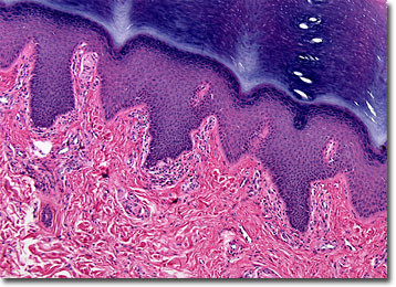
Similar to fingerprints, the patterns that appear on the integument of the palms, generally referred to as palmar skin, are unique and may be used as a means of identification. The practical purpose of the tiny wrinkles and folds along the palms, however, is to facilitate gripping and to enhance tactile sensitivity.
View an image of the palmar skin section at 20x magnification.
In addition to being hairless and packed with sweat glands, palmar skin is relatively thick and keratinized. Indeed, the skin that lines the palms of a human is typically 0.8 to 1.4 millimeters thick, while most other parts of the body are only protected by an integument 0.1 millimeters thick. Within palmar skin, five morphologically discrete layers of tissue exist. The outermost layer is the stratum corneum, which is predominantly comprised of dead cells that are almost continuously sloughed from the body. As these cells are lost, new ones originate via mitosis from the innermost layer of palmar skin, the stratum basale, and gradually migrate through the other strata until they too are eventually exfoliated and replaced.
Palmar skin is attached to a sheet of tissue called the palmar fascia that underlies it by septa. This connection serves to help stabilize the skin, making the palms better suited for grasping various objects. Sometimes, however, the palmar fascia thickens, resulting in a condition known as Dupuytren’s disease. Symptoms of the condition include the appearance of nodules and pits in the skin of the palms, as well as a general thickening of the integument. In severe cases, thickened cord-like structures may also occur and extend into the fingers. Such changes in the hands may cause the fingers to turn inwards toward the palm. Though pain from the disease is unusual, it may limit the range of motion of the hands and fingers, worsening the longer the condition is left untreated.
BACK TO THE BRIGHTFIELD MICROSCOPY IMAGE GALLERY