
Brightfield Microscopy Digital Image Gallery
Keloid Scar Tissue
When skin tissue is injured, as through a cut or a burn, it naturally forms a scar during the healing process. Most scars diminish in size over time or disappear altogether.
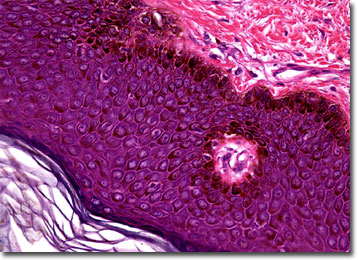
View an image of the keloid scar tissue section at 20x magnification.
Scar tissue is primarily composed of collagen, which is produced by fibroblasts in areas of the skin near the site of the wound. Typically the collagen does not exceed beyond the injured area of skin. However, some people form keloids when their skin is wounded, though scientists do not fully understand why this occurs. A keloid is a type of swollen scar that grows much larger than other scars, appearing similar to a fibrous tumor. Keloids are not especially dangerous, but they may cause discomfort and itching and may limit movement if they become too large, especially if they are located around the joints of the body. In some cases, they may be severely disfiguring as well.
Keloids form most often among young and dark skinned people, but others may also share a genetic disposition to form the overgrown scars. Similar to other types of scar tissue, keloids usually require medical treatment in order to improve their appearance, although there are documented cases of them diminishing in size on their own. However, not all people believe that keloids should be removed. In some cultures, the raised scars are created deliberately through controlled burning and cutting of the skin. This custom is often religious in nature and may serve as a rite of passage, signifying status of members within the group.
BACK TO THE BRIGHTFIELD MICROSCOPY IMAGE GALLERY